Copyright
©The Author(s) 2016.
World J Gastroenterol. Jan 21, 2016; 22(3): 1172-1178
Published online Jan 21, 2016. doi: 10.3748/wjg.v22.i3.1172
Published online Jan 21, 2016. doi: 10.3748/wjg.v22.i3.1172
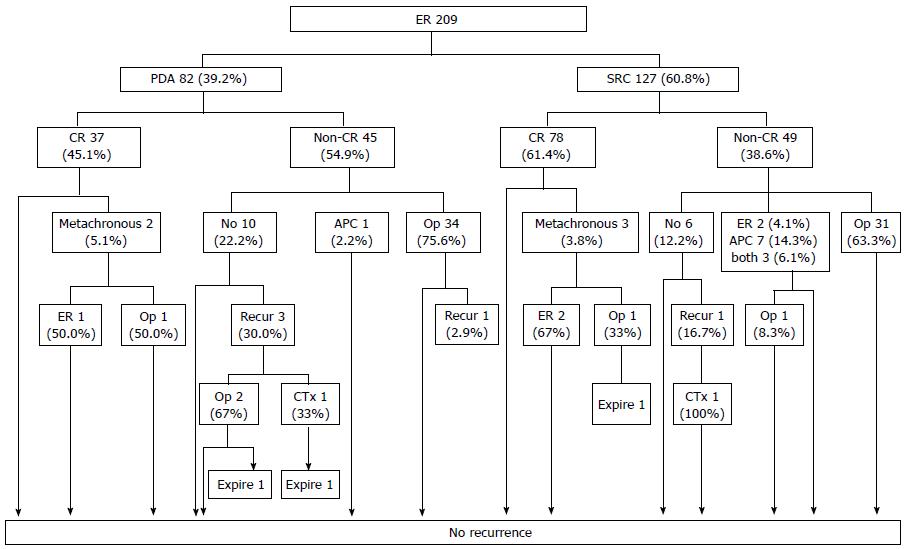
Figure 1 Follow-up outcomes after endoscopic resection of undifferentiated-type early gastric cancer.
The numbers in the boxes are the numbers of cases. CR: Curative resection; PDA: Poorly differentiated adenocarcinoma; SRC: Signet ring cell carcinoma; APC: Argon plasma coagulation; ER: Endoscopic resection. Taken with permission from Surg Endosc 2014; 28: 2627-2633[6].
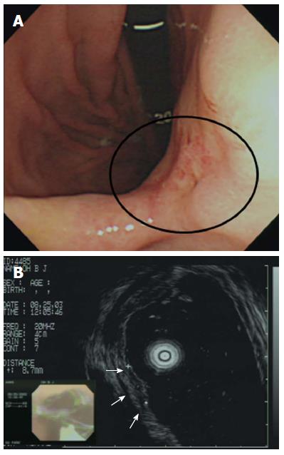
Figure 2 Incorrect T staging: a case of undifferentiated-type early gastric cancer (EGC).
A: Endoscopic image of an EGC showing a 10-mm-diameter depressed lesion in the posterior angle of the wall; B: Endoscopic ultrasonographic image showing a hypoechoic mucosal mass with an intact submucosal layer. A surgical specimen obtained upon radical subtotal gastrectomy confirmed that the EGC was confined to the submucosal layer. Taken with permission from Gastrointest Endosc 2007; 66: 901-908[35].
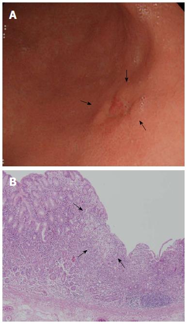
Figure 3 Clinical example of signet ring cell carcinoma exhibiting expansive intramucosal spread.
A: An endoscopic image of early gastric cancer (EGC) showing a depressed lesion located in the posterior wall of the lower body. Endoscopically, the surrounding mucosa did not exhibit atrophy or intestinal metaplasia; B: Pathological findings after endoscopic resection (× 40). SRC tumor cells were superficially exposed in a mucosal region and were well-demarcated (arrow). Taken with permission from Gut Liver 2015; 9: 720-726[40].
Figure 4 Clinical example of the signet ring cell carcinoma associated with infiltrative intramucosal spread.
A: An endoscopic image of an EGC, showing a flat lesion located in the lesser curvature of the lower body (circle). Endoscopically, the surrounding mucosa exhibited atrophic gastritis; B: After endoscopic resection, three of the lateral margins were positive (circle); C: Pathological findings after endoscopic resection (× 40). Signet ring cell carcinoma cells exhibited subepithelial spread. In other words, the lesion was of the infiltrative type (circle). Taken with permission from Gut Liver 2015; 9: 720-726[40].
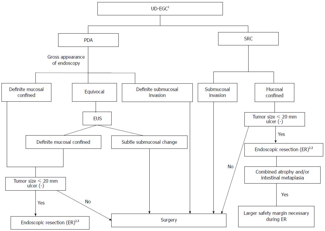
Figure 5 Suggested decision algorithm for endoscopic resection of undifferentiated-type early gastric cancer.
1Biopsy of several peripheral regions may aid in the exact diagnosis of undifferentiated-type histology prior to ER; 2Histologically minimum lateral margins should be wider than 3 mm for curative resection after ER; 3Even when complete resection has been achieved, short-term follow-up endoscopy to detect undifferentiated histology after ER may help to evaluate the risk of residual tumor development. PDA: Poorly differentiated adenocarcinoma; SRC: Signet ring cell carcinoma; EUS: Endoscopic ultrasonography; ER: Endoscopic resection; UD-EGC: Undifferentiated-type early gastric cancer.
- Citation: Kim JH. Important considerations when contemplating endoscopic resection of undifferentiated-type early gastric cancer. World J Gastroenterol 2016; 22(3): 1172-1178
- URL: https://www.wjgnet.com/1007-9327/full/v22/i3/1172.htm
- DOI: https://dx.doi.org/10.3748/wjg.v22.i3.1172


