©2013 Baishideng Publishing Group Co.
World J Gastroenterol. Aug 21, 2013; 19(31): 5061-5066
Published online Aug 21, 2013. doi: 10.3748/wjg.v19.i31.5061
Published online Aug 21, 2013. doi: 10.3748/wjg.v19.i31.5061
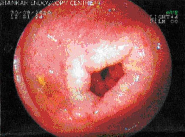
Figure 1 Endoscopy showing small superficial ulcers in stomach.
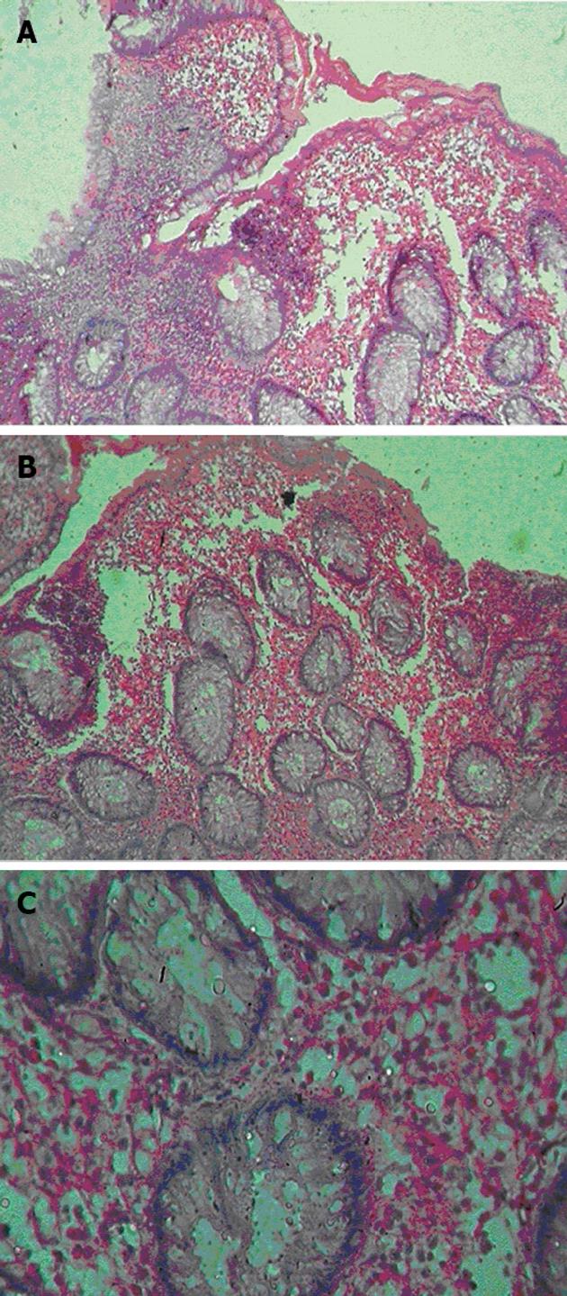
Figure 2 Large numbers of eosinophils are often present in the muscularis and serosa.
A, B: Showing dense eosinophilic infiltrates in the lamina propria and mucosa (× 10); C: Showing dense eosinophilic infiltrates in the lamina propria and mucosa (× 40).
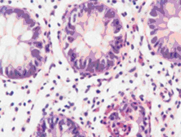
Figure 3 Post treatment (low dose steroid) biopsy showing resolution of disease.
- Citation: Ingle SB, Hinge (Ingle) CR. Eosinophilic gastroenteritis: An unusual type of gastroenteritis. World J Gastroenterol 2013; 19(31): 5061-5066
- URL: https://www.wjgnet.com/1007-9327/full/v19/i31/5061.htm
- DOI: https://dx.doi.org/10.3748/wjg.v19.i31.5061


