©2012 Baishideng Publishing Group Co.
World J Gastroenterol. Nov 21, 2012; 18(43): 6263-6268
Published online Nov 21, 2012. doi: 10.3748/wjg.v18.i43.6263
Published online Nov 21, 2012. doi: 10.3748/wjg.v18.i43.6263
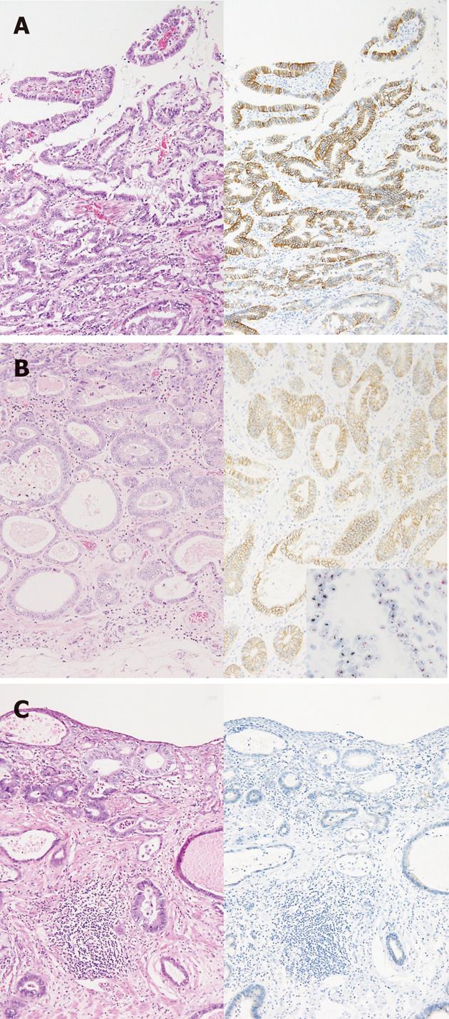
Figure 1 Immunohistochemical pattern of Barrett’s adenocarcinoma.
A: Immunohistochemistry (IHC) 3+ reaction in a well-differentiated tumor with papillary growth pattern; B: IHC 2+ reaction in a well-differentiated tumor with tubular pattern. Human epidermal growth factor receptor 2 (HER2) dual-color in situ hybridization amplification HER2/ centromeric enumeration probe ratio = 3.18; C: IHC 0 reaction in a well-differentiated tumor.
- Citation: Tanaka T, Fujimura A, Ichimura K, Yanai H, Sato Y, Takata K, Okada H, Kawano S, Tanabe S, Yoshino T. Clinicopathological characteristics of human epidermal growth factor receptor 2-positive Barrett's adenocarcinoma. World J Gastroenterol 2012; 18(43): 6263-6268
- URL: https://www.wjgnet.com/1007-9327/full/v18/i43/6263.htm
- DOI: https://dx.doi.org/10.3748/wjg.v18.i43.6263


