©2012 Baishideng Publishing Group Co.
World J Gastroenterol. Oct 21, 2012; 18(39): 5645-5648
Published online Oct 21, 2012. doi: 10.3748/wjg.v18.i39.5645
Published online Oct 21, 2012. doi: 10.3748/wjg.v18.i39.5645
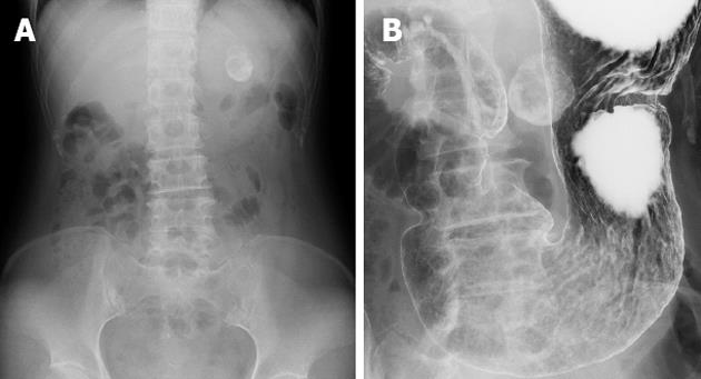
Figure 1 Radiological findings.
A: Abdominal X-ray indicated a spherical calcified lesion in the left upper quadrant; B: Barium study showed a round calcification bordering the gastric wall.
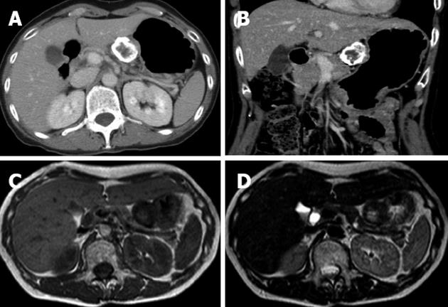
Figure 2 Computed tomography and magnetic resonance imaging findings.
Contrast-enhanced axial (A) and coronal (B) computed tomography examination demonstrated that the marginal zone of the tumor was calcified, and that the internal portion of the tumor was enhanced heterogeneously. Magnetic resonance imaging T1-weighted image (C) and T2-weighted image (D) revealed a low intensity marginal zone of tumor reflecting calcification.
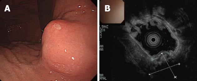
Figure 3 Endoscopy and endoscopic ultrasound of stomach.
A: Endoscopic examination revealed a round submucosal tumor; B: The tumor originated from the fourth layer of the gastric wall as indicated by endoscopic ultrasound. The deeper section could not be visualized because of calcification.
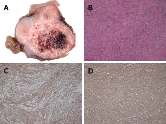
Figure 4 Pathological findings.
A: Sliced sections of the resected mass demonstrated a firm, solid, whitish-gray parenchyma with circular calcification and internal bleeding; B: Microscopically, the tumor was characterized by spindle-shaped tumor cells (hematoxylin and eosin, original magnification ×100); Immunohistochemically, the tumor cells were positive for KIT (C) and CD34 (D).
- Citation: Izawa N, Sawada T, Abiko R, Kumon D, Hirakawa M, Kobayashi M, Obinata N, Nomoto M, Maehata T, Yamauchi SI, Kouro T, Tsuda T, Kitajima S, Yasuda H, Tanaka K, Tanaka I, Hoshikawa M, Takagi M, Itoh F. Gastrointestinal stromal tumor presenting with prominent calcification. World J Gastroenterol 2012; 18(39): 5645-5648
- URL: https://www.wjgnet.com/1007-9327/full/v18/i39/5645.htm
- DOI: https://dx.doi.org/10.3748/wjg.v18.i39.5645


