Copyright
©2008 The WJG Press and Baishideng.
World J Gastroenterol. Jun 28, 2008; 14(24): 3914-3918
Published online Jun 28, 2008. doi: 10.3748/wjg.14.3914
Published online Jun 28, 2008. doi: 10.3748/wjg.14.3914
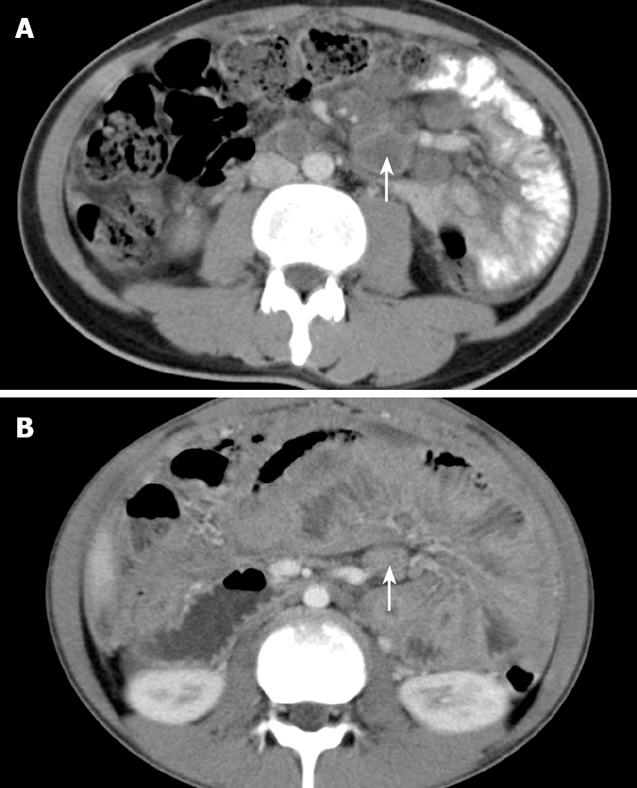
Figure 1 Contrast enhanced CT scan for a 25-year-old man with mesenteric TL showing enlarged lymph nodes in the body and root of SBM with peripheral enhancement (arrow) (A) and in the body of SBM with homogeneous enhancement (arrow) (B).
The SBM was contracted and the wall of the small bowel was thickened.
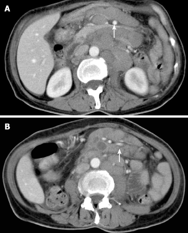
Figure 2 Contrast enhanced CT scan for a 56-year-old woman with NHL involving SBM showing enlarged lymph nodes in the root of SBM encasing the superior mesenteric artery (arrow), producing the “sandwich sign” (A) and homogeneously mixed peripheral enhancement of lymph nodes in the body of SBM encasing the small bowel mesenteric vessels (arrow), producing the “sandwich sign” (B).
-
Citation: Dong P, Wang B, Sun QY, Cui H. Tuberculosis
versus non-Hodgkin’s lymphomas involving small bowel mesentery: Evaluation with contrast-enhanced computed tomography. World J Gastroenterol 2008; 14(24): 3914-3918 - URL: https://www.wjgnet.com/1007-9327/full/v14/i24/3914.htm
- DOI: https://dx.doi.org/10.3748/wjg.14.3914


