Copyright
©2007 Baishideng Publishing Group Co.
World J Gastroenterol. Aug 28, 2007; 13(32): 4401-4404
Published online Aug 28, 2007. doi: 10.3748/wjg.v13.i32.4401
Published online Aug 28, 2007. doi: 10.3748/wjg.v13.i32.4401
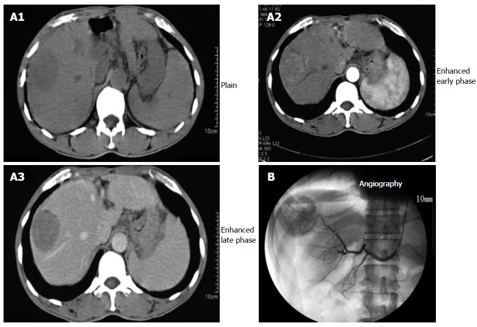
Figure 1 Helical dynamic abdominal CT revealing a liver tumor with a maximum diameter of 5.
2 cm in segment 8 (A) and abdominal angiography showing hyperva-scularity in it (B).
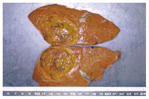
Figure 2 Gross appearance of resected specimen.
The cut surface of the resected liver specimen (S8) showed an encapsulated yellow-brownish tumor measuring 52 mm × 40 mm and 250 g.
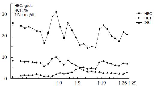
Figure 3 Postoperative course.
↑0: Operation, ↑9:Fever up, ↑19: Sleep out, ↑26: Blood transfusion of washed blood cells, ↑29: Discharged and started intake of lamivudine.
- Citation: Okada T, Kubota K, Kita J, Kato M, Sawada T. Hepatocellular carcinoma with chronic B-type hepatitis complicated by autoimmune hemolytic anemia: A case report. World J Gastroenterol 2007; 13(32): 4401-4404
- URL: https://www.wjgnet.com/1007-9327/full/v13/i32/4401.htm
- DOI: https://dx.doi.org/10.3748/wjg.v13.i32.4401


