©2006 Baishideng Publishing Group Co.
World J Gastroenterol. Apr 7, 2006; 12(13): 2136-2138
Published online Apr 7, 2006. doi: 10.3748/wjg.v12.i13.2136
Published online Apr 7, 2006. doi: 10.3748/wjg.v12.i13.2136
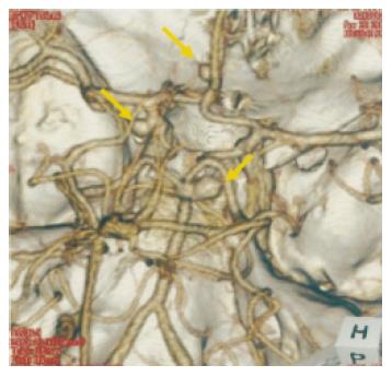
Figure 1 Computed tomographic angiography shows aneurysms of the left.
internal carotid-posterior communicating artery, left. anterior communicating artery, and right. posterior cerebral artery (arrows).
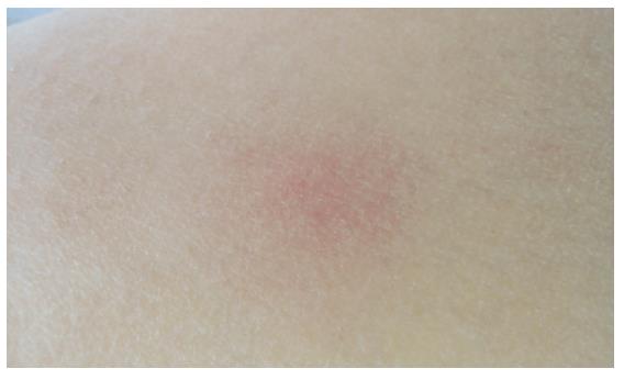
Figure 2 Typical eruption of erythema nodosum shows erythematous nodules on the anterior aspect of the leg.
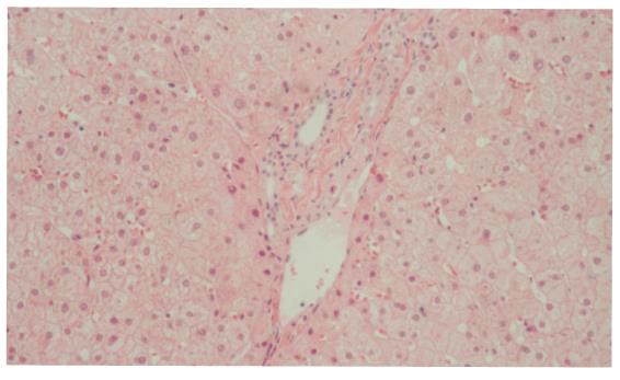
Figure 3 Liver biopsy specimen shows slightly enlarged portal tracts with no evidence of chronic nonsuppurative destructive cholangitis.
Hematoxylin and eosin, original magnification ×100.
- Citation: Iwadate H, Ohira H, Saito H, Takahashi A, Rai T, Takiguchi J, Sasajima T, Kobayashi H, Watanabe H, Sato Y. A case of primary biliary cirrhosis complicated by Behçet’s disease and palmoplantar pustulosis. World J Gastroenterol 2006; 12(13): 2136-2138
- URL: https://www.wjgnet.com/1007-9327/full/v12/i13/2136.htm
- DOI: https://dx.doi.org/10.3748/wjg.v12.i13.2136


