Copyright
©2006 Baishideng Publishing Group Co.
World J Gastroenterol. Mar 14, 2006; 12(10): 1577-1582
Published online Mar 14, 2006. doi: 10.3748/wjg.v12.i10.1577
Published online Mar 14, 2006. doi: 10.3748/wjg.v12.i10.1577
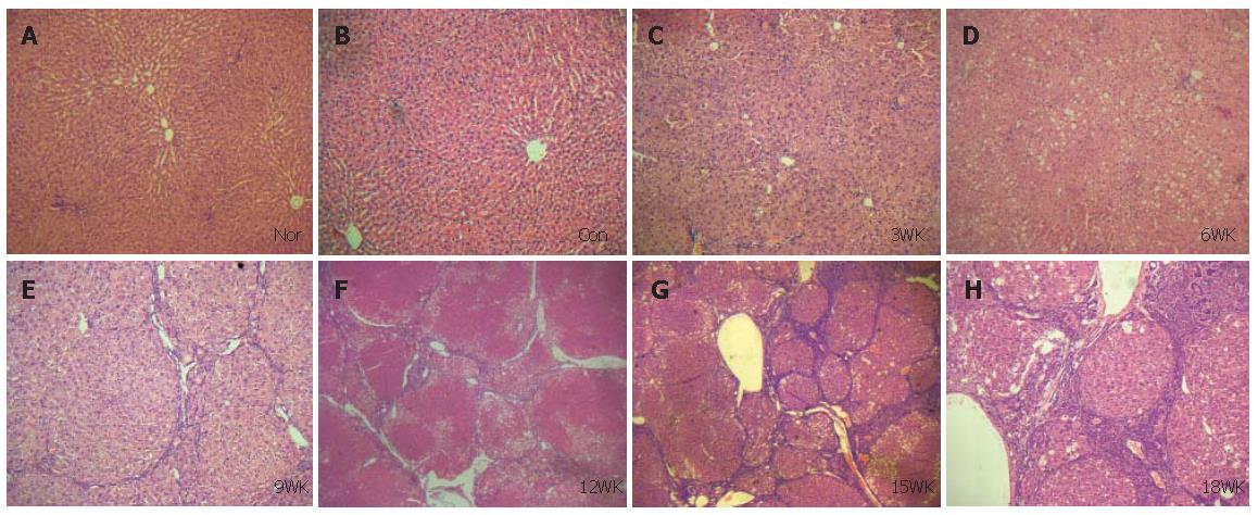
Figure 1 Pathology observation of the experimental rat liver sections stained with hematoxylin and eosin (×100).
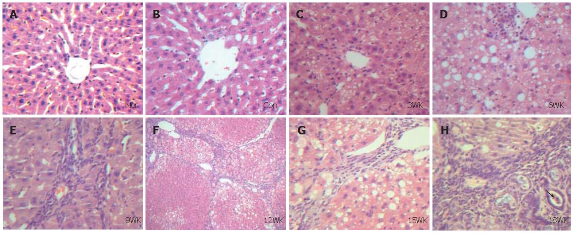
A: normal rats; B: control rats; C: CCl4 treated rats at 3 wk; D: CCl4 treated rats at 6 wk; E: CCl4 treated rats at 9 wk; F: CCl4 treated rats at 12 wk; G: CCl4 treated rats at 15 wk; H: CCl4 treated rats at 18 wk.
Figure 2 Pathology observation of the experimental rat liver sections stained with hematoxylin and eosin (×400).
The arrow shows atypical hyperplasia in the epithelium of bile capillary. A, B, C, D, E, F, G, H, see Figure 1.
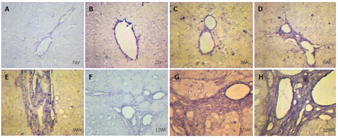
Figure 3 Expression of Smad2 mRNA in experimental rats liver sections during and after fibrosis (in situ hybridization ×400).
A, B, C, D, E, F, G, H, see Figure 1.
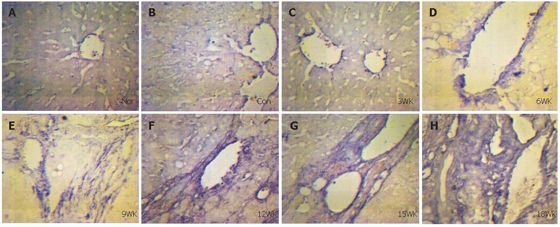
Figure 4 Expression of Smad4 mRNA in experimental rats liver sections during and after fibrosis (in situ hybridization ×400).
A, B, C, D, E, F, G, H, see Figure 1.
- Citation: Liu Y, Wang LF, Zou HF, Song XY, Xu HF, Lin P, Zheng HH, Yu XG. Expression and location of Smad2, 4 mRNAs during and after liver fibrogenesis of rats. World J Gastroenterol 2006; 12(10): 1577-1582
- URL: https://www.wjgnet.com/1007-9327/full/v12/i10/1577.htm
- DOI: https://dx.doi.org/10.3748/wjg.v12.i10.1577


