Published online Jan 26, 2021. doi: 10.12998/wjcc.v9.i3.690
Peer-review started: October 12, 2020
First decision: October 27, 2020
Revised: November 6, 2020
Accepted: November 29, 2020
Article in press: November 29, 2020
Published online: January 26, 2021
Processing time: 99 Days and 22 Hours
Currently, there have been no reports on foreign bodies found in the nasal septum after dental root canal therapy. Herein, we present an unusual case of a foreign body found in the nasal septum, which occurred after dental root canal therapy and two unsuccessful surgeries.
A 55-year-old man was referred to our department due to slight nasal discomfort that persisted for about 1 wk. Before consulting our department, the patient visited three different hospitals/clinics and underwent two surgeries that were not successful in removing a foreign body completely. A computed tomography scan was performed to detect the shift of the foreign body from dental root to the nasal septum, which resulted in the healing of oral inflammation and nasal septum discomfort. An endoscopic foreign body extraction surgery (3rd removal surgery) was then successfully performed, using a needle as the reference. No nasal reconstruction was required after the operation. Postoperative healing was uneventful.
Medical healthcare professionals should consider past medical history when dealing with foreign body cases. During septal foreign body extraction surgery, a needle could be used as a helpful reference.
Core Tip: In this case report, we present the case of a patient who visited four hospitals/clinics and underwent three surgical procedures for complete removal of a foreign body left during the dental root canal therapy. We consider this case suitable for publication for the following reasons: (1) This is an extremely unusual case. To the best of our knowledge, this is the first report on multiple nasal septal foreign bodies as a complication of dental root canal therapy; (2) It has educational value. This malpractice case calls for integrity reinforcement across medical institutions, as the foreign body left from the dental root canal therapy was deliberately covered up by the dentist until the foreign body broke. The patient visited four different hospitals/clinics and underwent three surgical procedures until the foreign bodies were totally removed; and (3) It has clinical value as it may provide an answer to the following questions: What should a dentist do when surgical instruments fracture? How to tell if there are two segments of a needle or if there is only one needle and the other segment is an artifact? Which surgical approach is the best for the patient?
- Citation: Du XW, Zhang JB, Xiao SF. Nasal septal foreign body as a complication of dental root canal therapy: A case report. World J Clin Cases 2021; 9(3): 690-696
- URL: https://www.wjgnet.com/2307-8960/full/v9/i3/690.htm
- DOI: https://dx.doi.org/10.12998/wjcc.v9.i3.690
After dental root canal therapy, foreign bodies may end up in the nasal septum. In most cases, these materials enter the nasal septum via oroantral communication, while in some rare cases, they enter via tooth socket or via the pulp chamber of a tooth. It is suspected that the septal foreign body found after dental root canal therapy occurs following needle tip migration. If not timely treated, it can cause septal abscess and loss of a part of the septal cartilage. Herein, we present an unusual case of a foreign body in the septum, which occurred after dental root canal therapy and two unsuccessful surgeries. We describe the patient’s course of events and treatment procedure, including his clinical and imaging findings.
A 55-year-old man was referred to the otorhinolaryngology department due to slight nasal discomfort that persisted for about 1 wk (timeline presented in Table 1).
| Event | Timeline |
| Dental root canal therapy (position 21) in clinic A | -4 wk |
| Swelling and pain in the upper gum and lip | -4 wk +1 d |
| Foreign bodies found at position 21 in clinic B | -3 wk |
| Dental root foreign body removal surgery in hospital C | -2 wk |
| Foreign body residue found, and the second removal surgery | -2 wk +1 d |
| Slight nasal discomfort | -1 wk |
| Presentation at our department | 0 d |
| Foreign bodies found in the nasal septum | +1 d |
| Nasal septal foreign body removal surgery | +2 d |
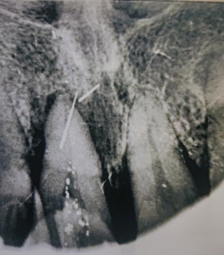
Four weeks before the consultation, the patient underwent dental root canal therapy (position 21) in clinic A. Two days after surgery, the patient had swelling and pain in the upper gum and lip, which were suspected to be caused by postoperative inflammation. He was then prescribed antibiotics and nonsteroidal antiinflammatory drugs. Yet, these problems persisted for 1 wk. The patient was then transferred to clinic B. A dental film was taken, revealing two hyperdense and pin-like masses at position 21 (Figure 1). The second dentist suspected a fishbone and referred the patient to hospital C. According to the dental film (Figure 1), the patient was diagnosed with a foreign body (the other hyperdense mass was an artifact). Consequently, a dental root foreign body removal surgery was performed under general anesthesia by labiogingival incision, and one piece of pin-like foreign body was successfully removed.
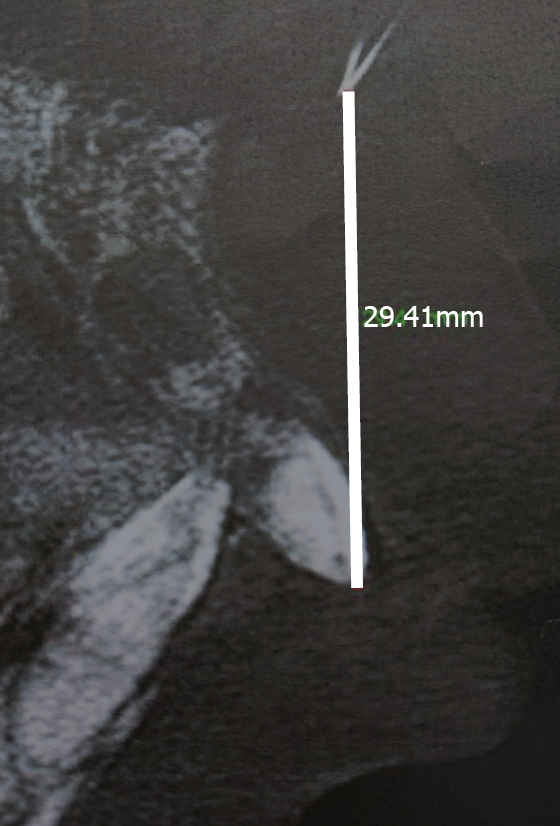
On the second day post-operation, the dental radiography was repeated, revealing a foreign body residue of 29.4 mm from the left incisor (Figure 2). A second foreign body removal surgery was performed under local anesthesia via original labiogingival incision; however, the foreign body was not found despite the long intervention.
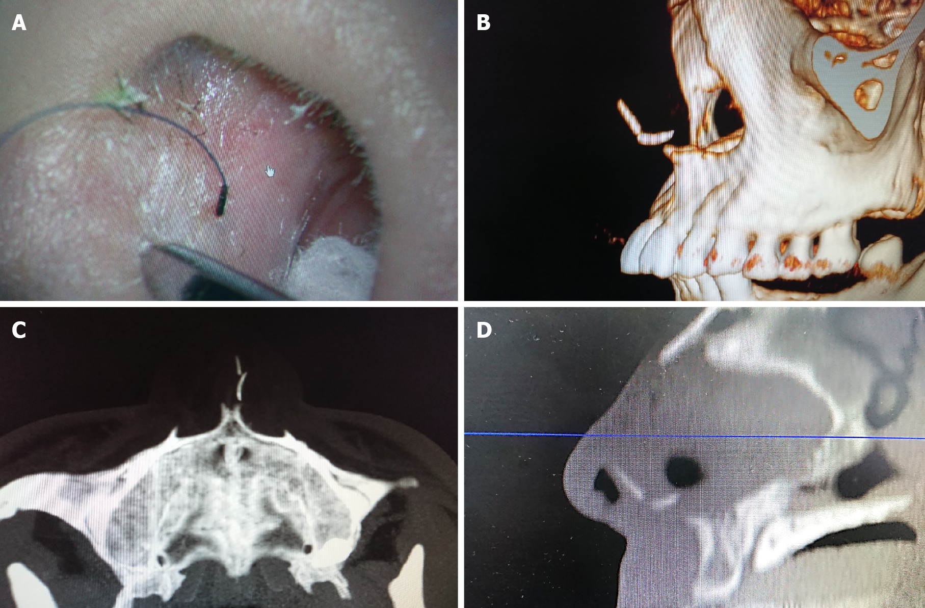
After the second surgery, the patient developed slight nasal discomfort that lasted for about 1 wk, after which the patient consulted our department. He underwent a computed tomography (CT) scan of the nasal sinuses. The axial (Figure 3A), sagittal (Figure 3B), and coronal (Figure 3C) planes showed a pin-like foreign body with hyperdensity in the left side of the nasal septum; three-dimensional reconstruction (Figure 3D) reconfirmed the position.
The patient was otherwise healthy and had no record of previous surgical interventions or the use of medication.
The patient had no relevant personal or family history.
The patient was immediately admitted. At the intraoral examination, granulation tissues were not evident in area 21. No sign of the foreign body was found following careful digital palpation, except for mild edema in the nasal septum.
The patient’s general condition was good, with normal hemoglobin levels and slightly elevated white blood cell count.
An emergency operation was scheduled. Before the septal foreign body extraction surgery, a needle was used as the reference (Figure 4A). A CT scan, which was performed to localize the foreign body and to avoid the same scenario from the last surgery, showed the needle pointing right to the pin-like foreign body (Figure 4B-D).
The diagnosis of a nasal septal foreign body was confirmed.
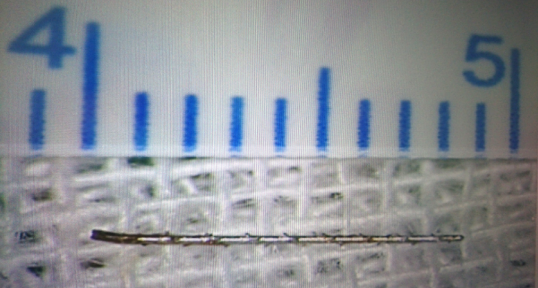
With the needle reference's help, the surgery was completed in half an hour under local anesthesia. The patient underwent surgical treatment for endoscopic extraction of a foreign body that was confirmed to be a piece of a fractured instrument for root canal therapy (Figure 5). The septal cartilage was intact, and no reconstruction was required. The patient was discharged from the hospital the following morning, medicated with a 7-d antibiotic treatment with cefdinir, analgesics, and nasal irrigation therapy. The recovery was uneventful.
At the 1-mo follow-up visit, the patient reported complete resolution of symptoms and did not exhibit septal abscess or deformity.
To the best of our knowledge, this is the first case report of a nasal septal foreign body following dental root canal therapy without recent nasal trauma or rhinoseptoplasty. An instrument fracturing inside a root canal is a relatively frequent and undesirable mishap during endodontic treatment[1]. Fractured instruments inside the root canal should be diagnosed, documented by an appropriate image examination, and recorded[2]. Clinical and radiological examinations performed by the maxillofacial surgeon could be used to rule out the odontogenic infection. Though it may seem that retained fractured instruments do not reduce the prognosis of endodontically treated teeth in the absence of apical periodontitis, healing may be significantly reduced if the disease is present[2]. Also, if an instrument fracture occurs, the patient should always be informed. The doctor can opt to leave the instrument inside the canal or remove it via the intracanal approach or surgery[2-4]. If surgery is needed, transnasal approaches should be preferred for retrieval of the foreign body[5].
Cases of foreign body migration into the paranasal sinus, orbit, and other adjacent craniofacial structures have been previously reported[6,7]. Additionally, some patients with implant migration have developed fungal infections and even cancer[8]. Foreign bodies in the sinuses should be surgically addressed. If it is complicated with abscess, the treatment should also include surgical drainage of the abscess and nasal packing in the acute phase, followed by antibiotic treatment. Because the infection and accumulation of pus between the cartilage and mucoperichondrium lead to ischemia and necrosis of the cartilage within 24 h, there is a high risk of septal cartilage destruction causing cartilaginous saddle nose deformation if not instantly treated.
The migration can occur due to surgical inexperience, insufficient planning, changes in intranasal pressure, autoimmune reactions, bone resorption, improper occlusal forces, and/or bone deficiencies[9-11]. In this case, a combination of several factors was probably responsible for this condition, including fracture of instruments inside a root canal and the subsequent surgeries. The route of the transposition of the foreign body from dental root to the nasal septum was assumed to be the following: Periodontal space → anterior space of the premaxillary bone → anterior nasal spine → nasal septum.
To avoid these inconveniences, dentists should be gentle in managing instruments, avoiding extreme angles, and keeping in mind the prevalence of instrument fractures, such as rotary nickel-titanium instruments (over stainless-steel hand), the third apical canals, and retreatment cases[12]. Also, when the endodontic instrument fracture happens, it should be immediately addressed. Definitive management should be based on a thorough knowledge of each treatment option's success rates, balanced against potential risks of removal or file retention[2]. Informing obligation must be seriously implemented, accompanied by appropriate record keeping. In addition, close follow-up is needed to check for the potential appearance of inflammation symptoms. Pain, redness, swelling, or purulent discharge requires medication and surgical treatment. Finally, thorough preoperative examination and operative exploration are essential if the remnant foreign body needs to be managed via the second procedure.
In summary, medical health care professionals should consider past medical history when dealing with a foreign body case. During septal foreign body removal endoscopic surgery, a needle as the reference could be very helpful. Also, CT imaging is crucial for an accurate diagnosis of entire foreign body fragments in the nasal cavity or sinuses.
| 1. | Ramos Brito AC, Verner FS, Junqueira RB, Yamasaki MC, Queiroz PM, Freitas DQ, Oliveira-Santos C. Detection of Fractured Endodontic Instruments in Root Canals: Comparison between Different Digital Radiography Systems and Cone-beam Computed Tomography. J Endod. 2017;43:544-549. [RCA] [PubMed] [DOI] [Full Text] [Cited by in Crossref: 20] [Cited by in RCA: 27] [Article Influence: 3.0] [Reference Citation Analysis (0)] |
| 2. | McGuigan MB, Louca C, Duncan HF. Clinical decision-making after endodontic instrument fracture. Br Dent J. 2013;214:395-400. [RCA] [PubMed] [DOI] [Full Text] [Cited by in Crossref: 43] [Cited by in RCA: 54] [Article Influence: 4.5] [Reference Citation Analysis (0)] |
| 3. | Saunders JL, Eleazer PD, Zhang P, Michalek S. Effect of a separated instrument on bacterial penetration of obturated root canals. J Endod. 2004;30:177-179. [RCA] [PubMed] [DOI] [Full Text] [Cited by in Crossref: 34] [Cited by in RCA: 34] [Article Influence: 1.5] [Reference Citation Analysis (0)] |
| 4. | Gandevivala A, Parekh B, Poplai G, Sayed A. Surgical removal of fractured endodontic instrument in the periapex of mandibular first molar. J Int Oral Health. 2014;6:85-88. [PubMed] |
| 5. | Levin M, Sommer DD. Endoscopic removal of ectopic sinonasal teeth: a systematic review. J Otolaryngol Head Neck Surg. 2019;48:30. [RCA] [PubMed] [DOI] [Full Text] [Full Text (PDF)] [Cited by in Crossref: 7] [Cited by in RCA: 14] [Article Influence: 2.0] [Reference Citation Analysis (0)] |
| 6. | González-García A, González-García J, Diniz-Freitas M, García-García A, Bullón P. Accidental displacement and migration of endosseous implants into adjacent craniofacial structures: a review and update. Med Oral Patol Oral Cir Bucal. 2012;17:e769-e774. [RCA] [PubMed] [DOI] [Full Text] [Full Text (PDF)] [Cited by in Crossref: 44] [Cited by in RCA: 38] [Article Influence: 2.7] [Reference Citation Analysis (0)] |
| 7. | Sousa Menezes A, Costa NDRMD, Moreira FC, Ribeiro D. Incisive dental implant migration into the nasal septum. BMJ Case Rep. 2019;12. [RCA] [PubMed] [DOI] [Full Text] [Cited by in Crossref: 3] [Cited by in RCA: 10] [Article Influence: 1.4] [Reference Citation Analysis (0)] |
| 8. | Dundar S, Karlidag T, Keles E. Endoscopic Removal of a Dental Implant from Maxillary Sinus. J Craniofac Surg. 2017;28:1003-1004. [RCA] [PubMed] [DOI] [Full Text] [Cited by in Crossref: 12] [Cited by in RCA: 14] [Article Influence: 1.8] [Reference Citation Analysis (0)] |
| 9. | Eltas A, Dundar S, Eltas SD, Altun O, Yolcu U, Saybak A. Accidental Displacement of Dental Implants Into Both Maxillary Sinuses During Surgery. J Oral Implantol. 2015;41:601-603. [RCA] [PubMed] [DOI] [Full Text] [Cited by in Crossref: 9] [Cited by in RCA: 10] [Article Influence: 0.8] [Reference Citation Analysis (0)] |
| 10. | Galindo-Moreno P, Padial-Molina M, Avila G, Rios HF, Hernández-Cortés P, Wang HL. Complications associated with implant migration into the maxillary sinus cavity. Clin Oral Implants Res. 2012;23:1152-1160. [RCA] [PubMed] [DOI] [Full Text] [Cited by in Crossref: 45] [Cited by in RCA: 43] [Article Influence: 2.9] [Reference Citation Analysis (0)] |
| 11. | Nakajima Y, Omori M, Inoue K, Nakano H, Fukui N, Kato-Kogoe N, Yamamoto K, Suwa Y, Sunano A, Ueno T. Removal of a Fixation Screw That Was Forced Into the Postsuperior Maxillary Sinus Wall: Case Report. Implant Dent. 2019;28:313-316. [RCA] [PubMed] [DOI] [Full Text] [Cited by in Crossref: 2] [Cited by in RCA: 2] [Article Influence: 0.3] [Reference Citation Analysis (0)] |
| 12. | Tzanetakis GN, Kontakiotis EG, Maurikou DV, Marzelou MP. Prevalence and management of instrument fracture in the postgraduate endodontic program at the Dental School of Athens: a five-year retrospective clinical study. J Endod. 2008;34:675-678. [RCA] [PubMed] [DOI] [Full Text] [Cited by in Crossref: 61] [Cited by in RCA: 78] [Article Influence: 4.3] [Reference Citation Analysis (1)] |
Open-Access: This article is an open-access article that was selected by an in-house editor and fully peer-reviewed by external reviewers. It is distributed in accordance with the Creative Commons Attribution NonCommercial (CC BY-NC 4.0) license, which permits others to distribute, remix, adapt, build upon this work non-commercially, and license their derivative works on different terms, provided the original work is properly cited and the use is non-commercial. See: http://creativecommons.org/Licenses/by-nc/4.0/
Manuscript source: Unsolicited manuscript
Specialty type: Medicine, research and experimental
Country/Territory of origin: China
Peer-review report’s scientific quality classification
Grade A (Excellent): 0
Grade B (Very good): 0
Grade C (Good): C
Grade D (Fair): 0
Grade E (Poor): 0
P-Reviewer: Cianci P S-Editor: Zhang L L-Editor: Wang TQ P-Editor: Yuan YY


