Published online Apr 26, 2019. doi: 10.12998/wjcc.v7.i8.1001
Peer-review started: December 23, 2018
First decision: January 12, 2019
Revised: January 31, 2019
Accepted: February 18, 2019
Article in press: February 18, 2019
Published online: April 26, 2019
Processing time: 126 Days and 6.2 Hours
We present a rare case of plasma cell type of Castleman’s disease (CD) involving only the right renal sinus in a 65-year-old woman with a duplex collecting system (DCS).
The patient presented with a right renal sinus lesion after renal ultrasonography. Subsequent abdominal enhanced computed tomography (CT) and magnetic resonance imaging (MRI) of the kidneys showed DCS and a soft tissue mass with mild enhancement at the lower right renal sinus. The lesion was suspected to be a malignant renal pelvic carcinoma. Hence, the patient underwent a right radical nephrectomy. Histological examination revealed hyperplastic lymphoid follicles in the renal sinus. A detailed review of the patient’s CT and MRI images and a literature review suggested that the lesion was hypointense on T2-weighted images and hyperintense on diffusion-weighted image manifestations, and showed mild enhancement, which distinguished the plasma cell type of CD from many other renal sinus lesions. Furthermore, peripelvic soft tissue masses with a smooth internal surface of the renal pelvis were on imaging findings, which suggests that the urinary tract epithelial system is invulnerable and can be used to differentiate the plasma cell type of CD from malignant lymphoma with a focally growth pattern to some extent.
Preoperative diagnosis is often difficult in such cases, as plasma cell type of CD involving only the right kidney is exceedingly rare. However, heightened awareness of this disease entity and its radiographic presentations may alert one to consider this diagnosis.
Core tip: We describe a rare case of plasma cell type of Castleman’s disease (CD) involving only the right renal sinus in a 65-year-old woman with a duplex collecting system. The imaging findings and pathological features of CD involving the renal sinus are summarized in this literature review.
- Citation: Guo XW, Jia XD, Shen SS, Ji H, Chen YM, Du Q, Zhang SQ. Radiologic features of Castleman’s disease involving the renal sinus: A case report and review of the literature. World J Clin Cases 2019; 7(8): 1001-1005
- URL: https://www.wjgnet.com/2307-8960/full/v7/i8/1001.htm
- DOI: https://dx.doi.org/10.12998/wjcc.v7.i8.1001
Castleman’s disease (CD) is a rare benign lymphoproliferative disease, and its pathogenesis is still unclear. CD usually involves mediastinal lymph nodes. Extrathoracic presentations have rarely been described, with even fewer presentations in the kidneys. CD can be histologically classified into three types as follows: Hyaline vascular type, plasma cell type, and mixed type. Preoperative diagnosis by imaging examination is very difficult to differentiate the plasma cell type of CD from malignant lymphoma. Here, we report a case of plasma cell type CD with only renal sinuses involved. We hope to identify the imaging features that can help the preoperative qualitative diagnosis of this disease through a literature review. Our case is unique because it is the first report of CD presenting as sinus tumors in a patient with a duplex collecting system (DCS)
A 65-year-old woman showed right renal sinus lesions on renal ultrasonography performed as part of a healthy physical examination. Medical history was negative for symptoms of discomfort.
Her history was unremarkable.
Laboratory findings showed mild liver dysfunction. Urinalysis findings were within normal limits, and urine cytology showed no evidence of malignancy. No obvious abnormality was found in other laboratory examinations. Normal blood flow to the left kidney and slightly decreased blood flow to the right kidney were noted on nephro-dynamic images.
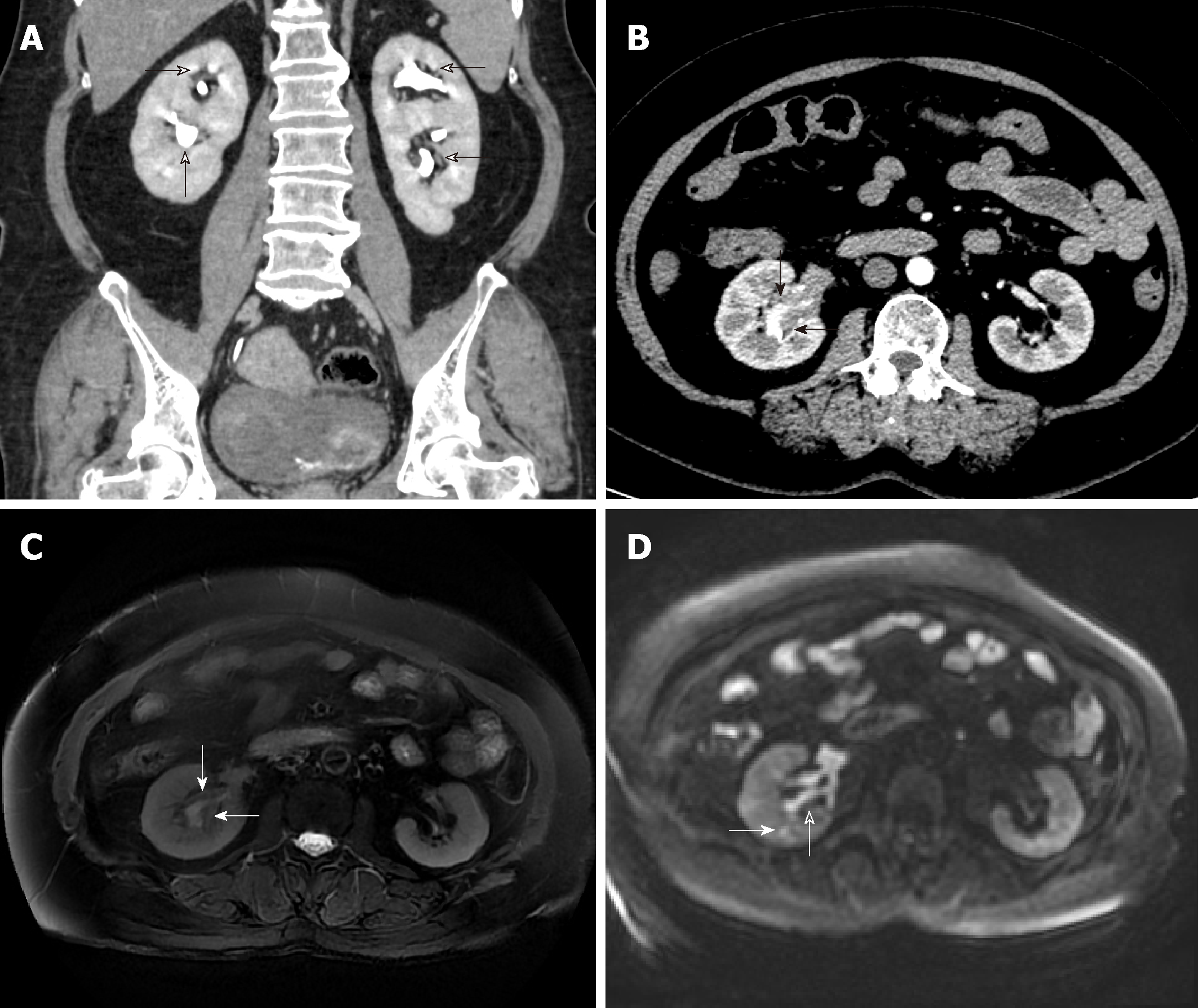
DCS and a soft tissue mass with mild enhancement at the lower right renal sinus were detected after subsequent abdominal computed tomography (CT). Magnetic resonance imaging (MRI) revealed a homogeneous mass that was isointense on M3D Lava in-phase images and hypointense on T2-weighted images (T2WI). Diffusion-weighted images (DWI) showed a hyperintense mass (Figure 1).
The final diagnosis was CD of plasma cell type (Figure 2).
The patient underwent a right radical nephrectomy. Histological examination revealed hyperplastic lymphoid follicles in the renal sinus and adjacent parenchyma. Immunohistochemical staining showed the following results: CD38 (+), CD138 (+), Kappa (+), Lambda (+), and CD21 (FDC+).
After treatment, thoracic and abdominal CT scans were used to determine whether the disease was multicentric. However, no enlarged lymph node or other lesions were found.
CD remains a rare and poorly understood disease characterized by massive growth of the lymphoid tissue. The renal sinus is an extremely unusual location for the manifestation of CD. The exact mechanism of CD in the renal sinus has not been reported thus far. Chronic low-grade inflammation, an immunodeficiency state, and autoimmunity have been proposed as probable mechanisms[1]. Patients with DCS are more susceptible to chronic inflammation, which is the probable cause of CD[2]. Our case supports chronic low-grade inflammation as a possible mechanism, although the exact cause of formation requires further study.
As far as we know, six articles on CD involving the renal sinus have been reported[3-8]. All lesions were confined to the renal sinus except in one case with a large range of lesions and retroperitoneal involvement. Six lesions in five cases were diagnosed as CD of plasma cell type, all of which were reported as case reports. Their imaging findings and pathological features are summarized in Table 1. In this table, the total number of patients with CD involvement of renal sinuses is six (seven lesions), including four males and two females, with an average age of 66 years. All of these patients presented homogeneous soft tissue density masses with mild enhancement, and the longest diameter less than 5 cm on CT image. Although the specific longest diameter of lesions was not given in some articles[4,5], the imaging data of lesions were given. Therefore, their size could be roughly obtained by comparison with the imaging data, in which the exact size of the lesions had been given. Four of these patients underwent MRI scans[4-6], showing isointense lesions on T1WI and hypointense lesions on T2WI, and two of these patients showed hyperintense lesions on DWI[4,5]. We believe that hypointensity on T2WI, hyperintensity on DWI manifestations, and mild enhancement can help us to distinguish the plasma cell type of CD from many renal sinus lesions, such as transitional cell carcinoma and renal sinus lymphoma. In most cases, the lesions of transitional cell carcinoma show high signal on T2WI, with unsmooth edges and hematuria as the most common clinical symptoms. However, it is still impossible to differentiate CD from renal sinus lymphoma by imaging alone. It also has lower signal intensity than a normal cortex with T1WI, and it is relatively iso- or hypointense in T2WI and hyperintense on DWI.
| Ref. | Jang et al[3] | Kin et al[4] | Nagahama et al[5] | Nishie et al[6] | Present | |
| Sex | M | M | M | M | F | F |
| Age (yr) | 64 | 59 | 79 | 70 | 65 | 62 |
| Sinus | Left | Left | Left | Bilateral | Unilateral | Right |
| MRI | ||||||
| T1WI | N/A | Isointense | Isointense | N/A | Isointense | Isointense |
| T2WI | N/A | Hypointense | Hypointense | N/A | Hypointense | Hypointense |
| DWI | N/A | Hyperintense | N/A | N/A | N/A | Hyperintense |
| CT size (cm) | 2.5-4 | N/A | N/A | 3.0-4.5 | 2.8-4.5 | |
| Enhancement | Mild | Mild | Mild | Mild | Mild | Mild |
| Clear boundary | Yes | Yes | Yes | Yes | Yes | Yes |
| Hydronephrosis | Mild | Mild | Mild | Mild | Mild | Mild |
| Urothelium intact | Yes | Yes | N/A | N/A | N/A | Yes |
In Table 1, only one of the seven lesions showed complete occlusion of the collecting system[5], and the rest of the lesions showed slight dilatation of renal pelvis and calyces. Therefore, compensatory dilatation of the collecting duct system was considered to be caused by subcutaneous lymphocyte hyperplasia in the urinary tract. The pathology of three cases clearly indicated that the pelvic tumor was a benign lymphoproliferative lesion with widely scattered, hyperplastic lymphoid follicles, while the pelvic urothelium was intact, which was consistent with the smooth and nonfilling defect on the inner surface of the collection system from CT and MRI imaging[3,4]. Sheth et al[9] reported that the imaging findings of renal lymphoma are related to the tumor growth pattern. If malignant lymphocytes proliferate focally, single or multiple well-defined masses can be formed and adjacent urinary tract systems destroyed. However, if it follows an infiltrative growth pattern, then the size of the kidney may increase without damage to the basic structure, and the lesions may be quite small with ill-defined borders. Similar well-defined renal sinus masses have been found in reported cases. If they are considered renal sinus lymphoma, then their growth mode should be focal growth, which is bound to be accompanied by adjacent urinary tract destruction. In fact, three cases showed complete urinary tract epithelium under an electron microscope. Although the remaining four cases failed to provide pathological evidence, these cases all shared similar imaging features, which show smooth intrarenal pelvic lateral surfaces. Therefore, we speculated that the plasma cell type of CD, due to its benign hyperplasia of lymphoid pathological, may not show epithelial invasion like lymphoma. We believe the imaging findings of the plasma cell type of CD, showing peripelvic soft tissue masses and a smooth internal surface of the renal pelvis, suggest that the urinary tract epithelial system is invulnerable. It can be used to differentiate the plasma cell type of CD from malignant lymphoma with a focal growth pattern. However, it is just a speculation and more cases are needed to verify it. Since the treatment of CD is different from that of lymphoma, the preoperative diagnosis of CD involving renal sinus will affect the choice of clinical treatment.
In conclusion, our case suggests that a patient with a DCS may have a risk of suffering CD due to his or her susceptibility to chronic inflammation, and imaging findings of the plasma cell type of CD, showing hypointensity on T2WI, hyperintensity on DWI, peripelvic soft tissue masses, and a smooth internal surface of the renal pelvis, are important to the preoperative diagnosis of CD of plasma cell type involving only the renal sinus.
Manuscript source: Unsolicited manuscript
Specialty type: Medicine, research and experimental
Country of origin: China
Peer-review report classification
Grade A (Excellent): 0
Grade B (Very good): B, B
Grade C (Good): C
Grade D (Fair): 0
Grade E (Poor): 0
P-Reviewer: Choi MR, Nechifor G, Ohashi N S-Editor: Ji FF L-Editor: Wang TQ E-Editor: Wu YXJ
| 1. | CASE RECORDS of the Massachusetts General Hospital: Case No. 40381. N Engl J Med. 1954;251:529-534. [RCA] [PubMed] [DOI] [Full Text] [Cited by in Crossref: 1] [Cited by in RCA: 1] [Article Influence: 0.0] [Reference Citation Analysis (0)] |
| 2. | Fernbach SK, Feinstein KA, Spencer K, Lindstrom CA. Ureteral duplication and its complications. Radiographics. 1997;17:109-127. [RCA] [PubMed] [DOI] [Full Text] [Cited by in Crossref: 110] [Cited by in RCA: 92] [Article Influence: 3.2] [Reference Citation Analysis (0)] |
| 3. | Jang SM, Han H, Jang KS, Jun YJ, Lee TY, Paik SS. Castleman's disease of the renal sinus presenting as a urothelial malignancy: a brief case report. Korean J Pathol. 2012;46:503-506. [RCA] [PubMed] [DOI] [Full Text] [Full Text (PDF)] [Cited by in Crossref: 3] [Cited by in RCA: 6] [Article Influence: 0.4] [Reference Citation Analysis (0)] |
| 4. | Kim TU, Kim S, Lee JW, Lee NK, Jeon UB, Ha HG, Shin DH. Plasma cell type of Castleman's disease involving renal parenchyma and sinus with cardiac tamponade: case report and literature review. Korean J Radiol. 2012;13:658-663. [RCA] [PubMed] [DOI] [Full Text] [Full Text (PDF)] [Cited by in Crossref: 9] [Cited by in RCA: 9] [Article Influence: 0.6] [Reference Citation Analysis (0)] |
| 5. | Nagahama K, Higashi K, Sanada S, Nezumi M, Itou H. [Multicentric Castleman's disease found by a renal sinus lesion: a case report]. Hinyokika Kiyo. 2000;46:95-99. [PubMed] |
| 6. | Nishie A, Yoshimitsu K, Irie H, Aibe H, Tajima T, Shinozaki K, Nakayama T, Kakihara D, Naito S, Ono M, Muranaka T, Honda H. Radiologic features of Castleman's disease occupying the renal sinus. AJR Am J Roentgenol. 2003;181:1037-1040. [RCA] [PubMed] [DOI] [Full Text] [Cited by in Crossref: 14] [Cited by in RCA: 13] [Article Influence: 0.6] [Reference Citation Analysis (0)] |
| 7. | Nolan RL, Banerjee A, Idikio H. Castleman's disease with vascular encasement and renal sinus involvement. Urol Radiol. 1988;10:173-175. [RCA] [PubMed] [DOI] [Full Text] [Cited by in Crossref: 6] [Cited by in RCA: 6] [Article Influence: 0.2] [Reference Citation Analysis (0)] |
| 8. | Park JB, Hwang JH, Kim H, Choe HS, Kim YK, Kim HB, Bang SM. Castleman disease presenting with jaundice: a case with the multicentric hyaline vascular variant. Korean J Intern Med. 2007;22:113-117. [RCA] [PubMed] [DOI] [Full Text] [Full Text (PDF)] [Cited by in Crossref: 22] [Cited by in RCA: 20] [Article Influence: 1.1] [Reference Citation Analysis (0)] |
| 9. | Sheth S, Ali S, Fishman E. Imaging of renal lymphoma: patterns of disease with pathologic correlation. Radiographics. 2006;26:1151-1168. [RCA] [PubMed] [DOI] [Full Text] [Cited by in Crossref: 147] [Cited by in RCA: 122] [Article Influence: 6.1] [Reference Citation Analysis (0)] |


