Published online Aug 16, 2014. doi: 10.12998/wjcc.v2.i8.377
Revised: May 22, 2014
Accepted: June 10, 2014
Published online: August 16, 2014
Processing time: 156 Days and 16.9 Hours
Renal cell carcinoma is a common urological malignancy with the unique ability to invade the inferior vena cava (IVC) and to extend into the right atrium of the heart. Of those with Renal cell carcinoma only 4%-25% are found to have IVC invasion and of those only 2%-10% extend into the right atrium. If treated surgically, extension of tumor thrombus is not a determinant of survival; therefore it is imperative to determine the presence and extent of tumor thrombus in order to determine surgical approach and tumor resection. To date this has been primarily accomplished by magnetic resonance imaging and computed tomography. We present a case of 61 years old African American woman in which transthoracic echocardiography provided a more accurate determination/characterization of the presence and degree of tumor thrombus and extension.
Core tip: Renal cell carcinoma is a common urological malignancy with the ability to invade the inferior vena cava and to extend into the right atrium of the heart. If treated surgically, extension of tumor thrombus is not a determinant of survival; therefore it is imperative to determine the presence and extent of tumor thrombus. To date, this has been primarily accomplished by magnetic resonance imaging and computed tomography; however, we present a case in which transthoracic echocardiography provided a more accurate determination/characterization of the presence and degree of tumor thrombus and extension.
- Citation: Bejarano M, Cameron YL, Koutlas TC, Movahed A. Transthoracic echo: A sensitive tool for detecting cardiac extension of renal cell carcinoma? World J Clin Cases 2014; 2(8): 377-379
- URL: https://www.wjgnet.com/2307-8960/full/v2/i8/377.htm
- DOI: https://dx.doi.org/10.12998/wjcc.v2.i8.377
Renal cell carcinoma is a common urological malignancy with the unique ability to invade the inferior vena cava (IVC) and to extend into the right atrium of the heart. Of those with renal cell carcinoma (RCC) only 4%-25% are found to have IVC invasion and of those only 2%-10% extend into the right atrium. If treated surgically, extension of tumor thrombus is not a determinant of survival; therefore it is imperative to determine the presence and extent of tumor thrombus in order to determine surgical approach and tumor resection. To date this has been primarily accomplished by magnetic resonance imaging (MRI) and computed tomography (CT).
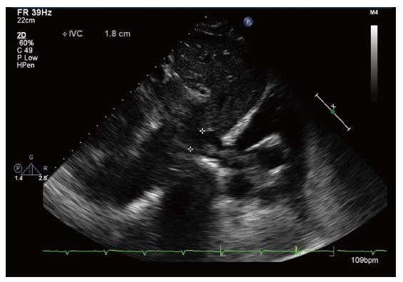
A 61 years old African-American female with past medical history of hypertension, hyperlipidemia, and diabetes mellitus initially presented to her primary care physician for progressively worsening fatigue, anorexia, and weight loss. As part of her work-up, she underwent an abdominal and pelvis CT scan, which revealed a large right sided renal mass with possible invasion of the right renal vein and inferior vena cava. A follow up abdominal MRI confirmed the presence of a very large right-sided renal mass consistent with renal cell carcinoma and subsequent invasion of the right renal vein and adjacent inferior vena cava. Further assessment revealed a tumor thrombus extending beyond the renal veins into the intrahepatic inferior vena cava and toward the right atrium. In order to fully characterize the extent of tumor thrombus extension, a CT angiogram (CTA) of the chest and transthoracic echocardiogram (TTE) was done. The chest CTA revealed a filling defect in the IVC in the intrahepatic and subhepatic regions consistent with known tumor thrombus, but showed no evidence of right atrium invasion (Figure 1). On the other hand, the TTE showed a large right atrial mass (Figure 2) with diastolic prolapse into the right ventricle and extension into the IVC (Figure 3). In the setting of her known history of RCC with migration and the fact that this mass was also seen to extend into the IVC, it was felt that this was indeed right atrial invasion of the tumor thrombus and less likely to be a hematological thrombus. A preoperative left heart catheterization was also performed revealing significant mid right coronary artery disease.
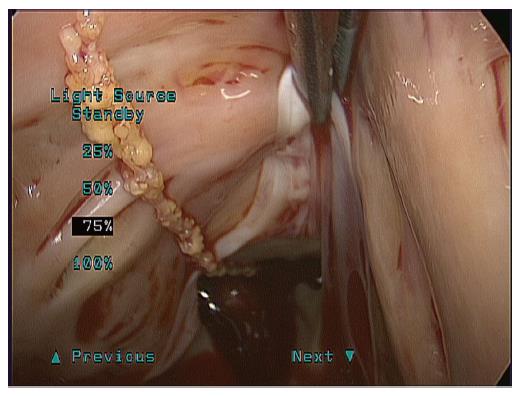
After completing the aforementioned preoperative assessment and evaluation by Urology, Vascular Surgery and Cardiothoracic Surgery, the patient underwent a radical nephrectomy and resection of the inferior vena cava and right atrial tumor thrombi (Figure 4). She simultaneously underwent a single vessel coronary artery bypass for her right coronary artery disease. There were no surgical complications and the patient’s postoperative course was unremarkable.
Renal cell carcinoma tumor thrombus has a propensity to invade the main renal veins as well as the IVC and in rare circumstances can extend into the right atrium of the heart[1,2]. In order to properly classify RCC extension and plan the appropriate surgical technique and approach, one would imagine that establishing the location of the superior margin of the tumor thrombus would be essential[1,3]. At this time the mainstay or gold standard of renal mass detection and characterization (including RCC) is CT scan and MRI[1,4,5]. CT scan has shown to be most accurate in evaluating the extent of local growth as well as the presence or absence of metastasis (i.e., to the pancreas, bone). On the other hand MRI has been more accurate in delineating the superior margin of any tumor thrombus, and thereby classifying RCC tumor thrombus, as well as differentiating between bland/hematologic thrombus and tumor thrombus[1,4,5]. Traditionally TTE has been used to further delineate the supradiaphragmatic extension of tumor thrombus. In our case, TTE accurately illustrated the cranial extent of tumor thrombus into the right atrium which was in fact missed on the traditionally used CT scan.
For level IV tumors (Table 1) such as was found in our patient, cardiopulmonary bypass (CPB) with or without hypothermic circulatory arrest (HCA) is necessary for safe and complete extraction of the thrombus[1,3,6]. This surgical approach provides a bloodless surgical field that allows optimal visualization of the hepatic veins, IVC and Right Atrium for complete tumor thrombus resection. As incomplete resection of these tumors confers a higher rate of metastatic recurrence and decreased postoperative survival, it is imperative to clearly delineate the superior margin of any tumor thrombus[1,3,6].
| Tumor thrombus level[6] | Characteristics[6] |
| Level I | Extension to 2 cm above the renal vein into the IVC |
| Level II | Extension to the subhepatic level, > 2 cm above the renal vein BUT below the diaphragm |
| Level III | Extension into the intrahepatic IVC BUT below the diaphragm |
| Level IV | Extension into the right atrium of the heart |
In our case, the patient’s preoperative evaluation included a CT abdomen/pelvis, CT chest, MRI abdomen, and TTE. Unexpectedly, it was the TTE that provided the most accurate determination of the cranial extent of the tumor thrombus. Proper classification of the tumor thrombus allowed for the appropriate surgical approach to be undertaken ensuring the best patient outcome.
A 61 years old African-American female with past medical history of hypertension, hyperlipidemia, and diabetes mellitus initially presented to her primary care physician for progressively worsening fatigue, anorexia, and weight loss.
A large right sided renal mass with possible invasion of the right renal vein and inferior vena cava.
Chest computed tomography angiogram revealed a filling defect in the inferior vena cava (IVC) in the intrahepatic and subhepatic regions consistent with known tumor thrombus, the transthoracic echocardiogram (TTE) showed a large right atrial mass with diastolic prolapse into the right ventricle and extension into the IVC.
In this case, TTE accurately illustrated the cranial extent of tumor thrombus into the right atrium which was in fact missed on the traditionally used computed tomography scan.
The case report illustrates the diagnostic power of transthoracic echo in diagnosis of cardiac extension of renal cell carcinoma. The manuscript is well written.
| 1. | Heidenreich A, Ravery V; European Society of Oncological Urology. Preoperative imaging in renal cell cancer. World J Urol. 2004;22:307-315. [RCA] [PubMed] [DOI] [Full Text] [Cited by in RCA: 1] [Reference Citation Analysis (0)] |
| 2. | Kallman DA, King BF, Hattery RR, Charboneau JW, Ehman RL, Guthman DA, Blute ML. Renal vein and inferior vena cava tumor thrombus in renal cell carcinoma: CT, US, MRI and venacavography. J Comput Assist Tomogr. 1992;16:240-247. [PubMed] |
| 3. | Posacioglu H, Ayik MF, Zeytunlu M, Amanvermez D, Engin C, Apaydin AZ. Management of renal cell carcinoma with intracardiac extension. J Card Surg. 2008;23:754-758. [RCA] [PubMed] [DOI] [Full Text] [Cited by in Crossref: 10] [Cited by in RCA: 10] [Article Influence: 0.6] [Reference Citation Analysis (0)] |
| 4. | Kang SK, Kim D, Chandarana H. Contemporary imaging of the renal mass. Curr Urol Rep. 2011;12:11-17. [RCA] [PubMed] [DOI] [Full Text] [Cited by in Crossref: 34] [Cited by in RCA: 31] [Article Influence: 2.1] [Reference Citation Analysis (0)] |
| 5. | Tollefson MK, Takahashi N, Leibovich BC. Contemporary imaging modalities for the surveillance of patients with renal cell carcinoma. Curr Urol Rep. 2007;8:38-43. [PubMed] |
| 6. | Boorjian SA, Sengupta S, Blute ML. Renal cell carcinoma: vena caval involvement. BJU Int. 2007;99:1239-1244. [RCA] [PubMed] [DOI] [Full Text] [Cited by in Crossref: 67] [Cited by in RCA: 69] [Article Influence: 3.6] [Reference Citation Analysis (0)] |
P- Reviewer: Chiu KW, Gassler N S- Editor: Song XX L- Editor: A E- Editor: Lu YJ


