Published online Oct 6, 2025. doi: 10.12998/wjcc.v13.i28.106852
Revised: April 20, 2025
Accepted: July 3, 2025
Published online: October 6, 2025
Processing time: 151 Days and 16.2 Hours
MURCS association, an acronym for Müllerian duct aplasia/hypoplasia, con
This report describes the successful anaesthetic and airway management of a 6-year-old girl with MURCS undergoing dental extraction. To address the comple
This report emphasizes individualized anesthetic strategies and interprofessional collaboration for managing rare congenital syndromes.
Core Tip: MURCS association, characterized by Müllerian duct aplasia, renal anomalies, and cervical spine dysplasia, poses significant airway management challenges due to spinal malformations. This report details the successful anesthetic management of a 6-year-old girl undergoing dental extraction. A multidisciplinary approach, including comprehensive preoperative assessment, meticulous planning, and advanced airway techniques, ensured procedural safety. The case highlights the importance of individualized anesthetic strategies and interprofessional collaboration to address airway complexities and other challenges in rare congenital syndromes, emphasizing the critical role of tailored management in achieving safe and successful outcomes.
- Citation: Lin XN, Chan WS, Lu CW. Airway management strategies in a pediatric patient with MURCS association: A case report. World J Clin Cases 2025; 13(28): 106852
- URL: https://www.wjgnet.com/2307-8960/full/v13/i28/106852.htm
- DOI: https://dx.doi.org/10.12998/wjcc.v13.i28.106852
MURCS is a rare congenital malformation complex comprising Müllerian duct aplasia/hypoplasia, renal agenesis/ectopy, and cervicothoracic somite dysplasia. It is considered a subtype of Mayer-Rokitansky-Küster-Hauser syndrome, typically affecting female patients and associated with short stature, scoliosis, and skeletal deformities involving the spine and upper limbs[1,2]. Cervical vertebral anomalies in MURCS can lead to restricted neck mobility, cervical spine instability, and potential brainstem compression, all of which complicate airway management and anaesthetic planning[3,4]. These anatomical abnormalities significantly increase the risk of neurological injury during airway manipulation, highlighting the importance of comprehensive preoperative evaluation and advanced airway strategies in affected patients. Despite its clinical implications, limited literature addressing the diagnosis, long-term outcomes, and anaesthetic management of MURCS patients.
Here, we report the case of a 6-year-old girl with MURCS who successfully underwent general anaesthesia for full-mouth dental extraction to treat severe caries.
A 6-year-and-1-month-old girl presented with multiple dental caries.
The patient had a history of MURCS association and exhibited short stature (height: 102 cm; weight: 13.7 kg; < 3rd percentile for age), amblyopia, congenital heart disease with an untreated bicuspid aortic valve, scapular anomalies, and thumb hypoplasia. She had received all age-appropriate vaccinations and she underwent regular outpatient follow-up for developmental therapy due to delayed developmental milestones.
The patient was referred to our hospital for further evaluation of the multiple dental caries. She was scheduled for dental surgery including tooth extraction, root canal treatment, and full-mouth rehabilitation. Owing to the lack of cooperation, general anaesthesia was recommended.
There is no family history of MURCS or other hereditary disorders. Previous surgical interventions included correction of scapular anomalies and thumb hypoplasia at another hospital.
Preoperative assessment revealed an adequate range of neck motion. Bilateral breath sounds were clear, and heart sounds were regular without murmurs. No specific abnormalities were detected on general physical examination.
Preoperative laboratory examinations, including complete blood count, electrolyte levels (potassium: 3.8 mmol/L, sodium, 137 mmol/L; chloride, 106 mmol/L), and liver function tests (alanine transaminase, 6 U/L), were within normal limits. However, a relatively low creatinine level (0.37 mg/dL) was noted on admission, whereas blood urea nitrogen (12 mg/dL), albumin (4.3 g/dL), and total protein (7.5 g/dL) levels remained within the normal range.
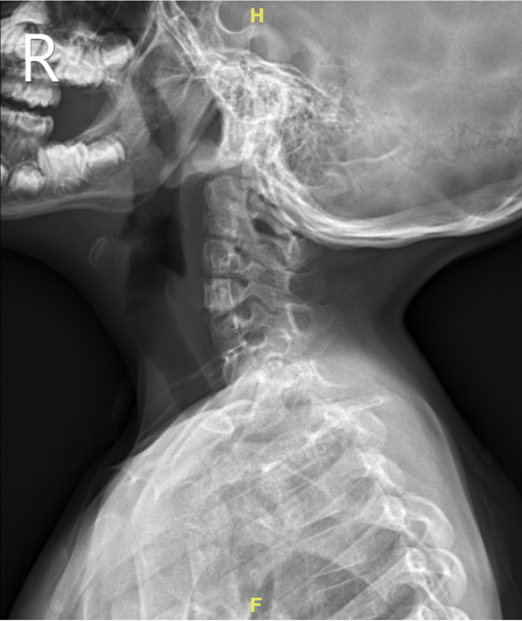
Preoperative echocardiography revealed a bicuspid aortic valve with trivial aortic regurgitation. Cervical spine radiography revealed a normal alignment without scoliosis or vertebral dysplasia (Figure 1). Abdominal ultrasonography was performed to exclude renal agenesis.
Dental caries status post complicated extraction, pulpotomy with endodontic therapy, and restorative treatment.
Following adequate disinfection and caries removal, pulpotomy was performed on teeth 54, 55, 64, 65, 74, 75, 84, and 85 under local anesthesia with 2% lidocaine and epinephrine (1:100000) and rubber dam isolation. Pulpotomy included hemostasis, filling with zinc oxide eugenol paste, and coronal sealing. Stainless steel crowns were subsequently placed on these teeth.
Root canal treatment and restorative procedures were performed on teeth 53, 62, and 63. Teeth 51, 52, and 61 were extracted using dental forceps and an elevator; subsequently gauze pack was applied. An intraoral periapical radiograph was obtained to evaluate outcomes, and postoperative medication was prescribed for infection control and pain management.
Upon arrival in the operating room, the patient was placed under American Society of Anaesthesiologists standard monitoring, including pulse oximetry, an oxygen analyser, capnography, electrocardiography, non-invasive blood pressure monitoring, and temperature monitoring. The initial heart rate was 112 bpm, and oxygen saturation was 99% on room air.
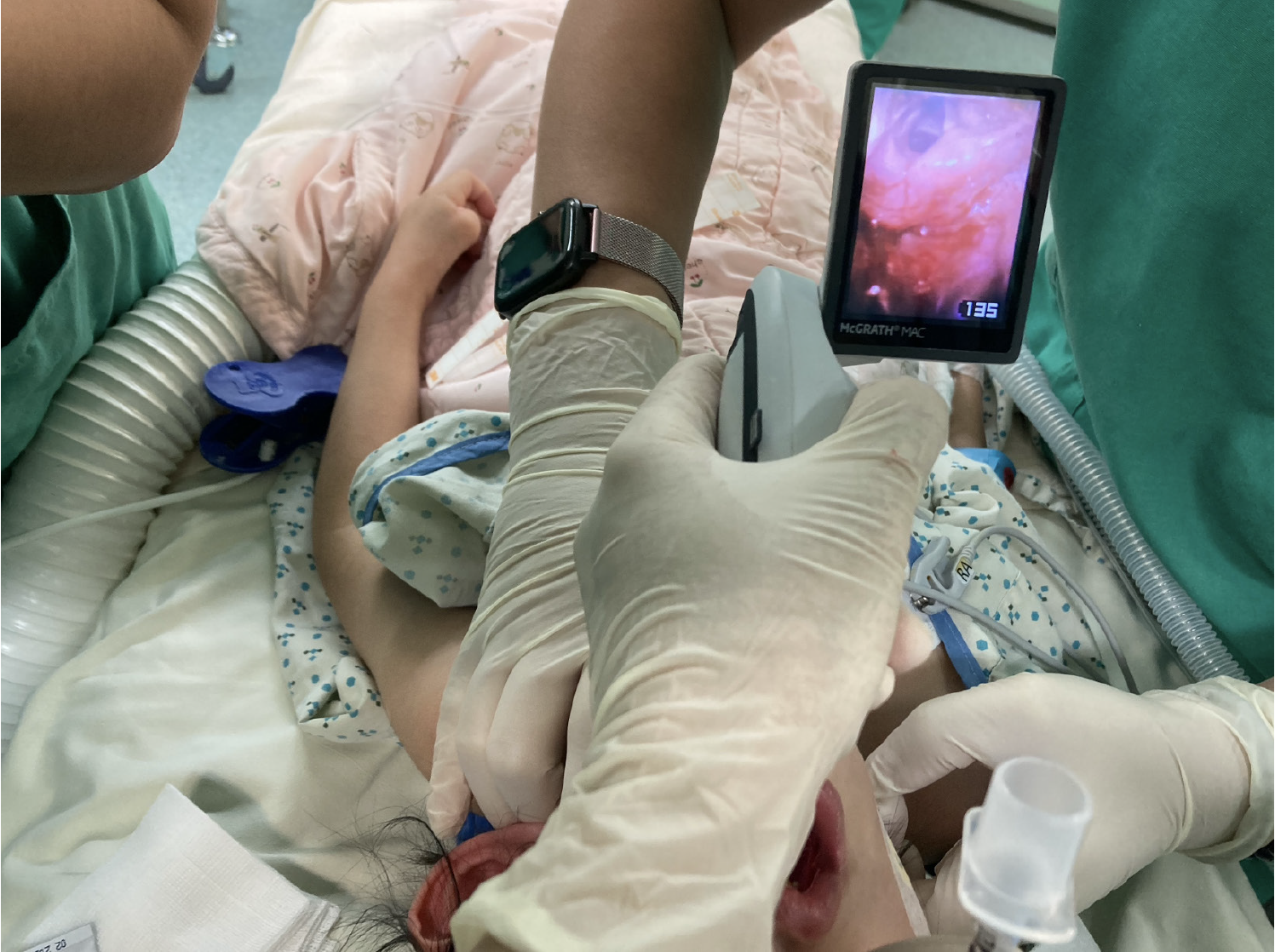
After preoxygenation, general anaesthesia was induced using sevoflurane inhalation and intravenous rocuronium (10 mg). A 4.5-mm endotracheal tube was used for nasal intubation and secured at a depth of 17 cm. Intubation was performed with a McGRATHTM MAC video laryngoscope using size 2 disposable blade. The procedure was relatively smooth, with a Cormack-Lehane classification grade of 2 on video laryngoscopy (Figure 2). No additional intraoperative rocuronium was administered.
The surgery lasted for 4 hours and 35 minutes and proceeded without complications. At the end of the procedure, sevoflurane was discontinued, and the neuromuscular blockade was reversed with sugammadex (27 mg, 2 mg/kg). Once the patient was able to open her eyes and demonstrate adequate spontaneous breathing (tidal volume approximately 110 mL), the endotracheal tube was removed without difficulty. Subsequently, the patient was transferred to the post-anaesthesia care unit (PACU) for continuous monitoring. Standard PACU monitoring, including pulse oximetry, electrocardiography, non-invasive blood pressure measurements, and temperature surveillance, was performed throughout the stay. Her parents accompanied her to provide reassurance and manage potential emergence agitation. No additional airway interventions were required, and the patient maintained adequate spontaneous ventilation with 100% oxygen saturation. After an uneventful 35-minute observation period, the patient was comfortably transferred to a general ward.
The patient was discharged uneventfully the following morning and was followed for one month postoperatively with no anaesthesia- or surgery-related sequelae.
Patients with MURCS association often present with complex craniovertebral and spinal anomalies, which may not always be apparent on plain radiographs but can substantially impact airway anatomy and anaesthetic risk. Cervical vertebral fusion, hypoplasia, or segmentation defects may limit neck extension, complicating direct laryngoscopy and increasing the risk of spinal cord injury[5,6]. Additionally, coexisting thoracic cage and scapular anomalies can further restrict positioning and ventilation. From an anaesthetic perspective, these patients require individualised airway assessment protocols. The use of video laryngoscopy, fibreoptic bronchoscopy, awake intubation should be considered. Risk stratification should also account for associated cardiac or renal anomalies, which are frequently observed in MURCS syndrome[2,7].
This case of a 6-year-old girl with MURCS association highlights the complexities of paediatric anaesthesia in the context of rare congenital anomalies. Her condition encompassed Müllerian duct aplasia, skeletal anomalies, a congenital cardiac valve defect, and developmental delay. Although a preoperative cervical spine radiograph showed normal alignment with no obvious vertebral malformations, no advanced imaging (computed tomography or magnetic reson
When formulating the anaesthetic plan, the team carefully accounted for patient comorbidities and unique risks. Maintaining haemodynamic stability was of paramount importance because of her congenital heart disease; therefore, agents and techniques with minimal cardiovascular depression were selected. Inhalational induction with sevoflurane was chosen because it allows slow and controlled induction and avoids the need for immediate intravenous access in an anxious, developmentally delayed child. The relatively gentle cardiovascular profile of sevoflurane helps prevent abrupt changes in heart rate or blood pressure, which is an important consideration for patients with valvular defects[8]. Preoxygenation was performed diligently before induction to maximise oxygen reserves, recognising that any difficulty in securing the airway could lengthen the apnoeic period. Additionally, the child’s developmental delay meant that cooperation would be limited. Mask induction with sevoflurane provided a calm transition to unconsciousness without the distress of awake IV placement or awake intubation attempts. This approach addresses both the psychological needs of paediatric patients and the clinical need to maintain stable cardiorespiratory conditions during induction.
Given the possibility of neuromuscular or musculoskeletal anomalies in patients with MURCS, muscle relaxation should be managed with caution. After the patient lost consciousness, a single dose of rocuronium (a non-depolarising neuromuscular blocker) was administered to facilitate intubation. Rocuronium was selected over a depolarising agent (such as succinylcholine) to avoid potential complications (e.g., hyperkalaemia in undiagnosed muscle disorders) and because it can be rapidly and reliably reversed[9]. No additional rocuronium was required during the 4.5-hour procedure, reflecting the strategy of using the minimal necessary paralytic. Neuromuscular function was continuously monitored intraoperatively to detect atypical responses or prolonged blockades. Importantly, the team was readily available and administered sugammadex (2 mg/kg at the end of the surgery) to fully reverse the neuromuscular block. Sugammadex provides a safety net against unanticipated prolonged paralysis, ensuring that patients regain adequate muscle strength for safe extubation[10]. By employing reversible muscle relaxation and maintaining vigilant monitoring, the anaesthesiology team mitigated the risk of neuromuscular anomalies or postoperative respiratory compromise in this patient.
A comprehensive airway strategy was implemented to avoid potential airway difficulties owing to MURCS-associated cervical spine abnormalities. Notably, the patient had no history of difficult intubation; however, known anatomical risk factors (possible cervical vertebral dysplasia) warrant preparation for a challenging airway. A range of advanced airway tools were prepared and immediately available at the bedside. This included a paediatric video laryngoscope (which was ultimately used), flexible fibreoptic bronchoscope, assorted sizes of supraglottic airway devices (laryngeal mask airways), and various endotracheal tube sizes. The plan also included adjuncts such as a stylet and Magill forceps for planned nasal intubation and readiness to perform an emergency surgical airway if required. The anaesthesia and surgical teams conducted a thorough briefing to ensure that each member was aware of the difficult airway plan, and roles were assigned when urgent intervention was necessary. By planning the worst-case scenario (could not intubate or ventilate), the team ensured that any escalation in airway management would be rapid and decisive.
Induction proceeded smoothly with these preparations. After anaesthesia induction with sevoflurane and rocuronium, nasal intubation was successfully achieved on the first attempt using a McGRATH™ MAC video laryngoscope with size 2 blade. The video laryngoscope provides an improved view of the glottis with minimal need for neck extension, which is crucial for protecting the cervical spine. The laryngoscopic view was Cormack-Lehane grade 2 (as visualised on the video screen), and a 4.5 mm cuffed endotracheal tube was passed without difficulty or the need for any intubation adjuncts. Manual inline stabilisation of the cervical spine was performed during intubation to ensure spinal stability. Intubation was performed without hypoxia, significant haemodynamic fluctuations, or trauma. Notably, no additional advanced techniques (such as fibreoptic intubation) or multiple attempts were required because the video laryngoscope approach proved sufficient. However, the presence of these backup devices was a key factor in the team’s confidence and safety margin. Ventilation and oxygenation remained adequate throughout the procedure and there were no signs of airway obstruction or bronchospasms. Careful planning and the use of video-assisted intubation in this case exemplified effective anticipation of difficulty and successful application of modern airway tools, resulting in a controlled and successful outcome, even in a high-risk scenario.
Despite the lack of obvious cervical spine deformity on radiography, patients with MURCS are at risk of complex airway challenges due to underlying skeletal abnormalities. Cervicothoracic somite dysplasia can manifest as subtle cervical vertebral fusion, instability, or malalignment that may not be evident without advanced imaging techniques. Thus, even in the absence of apparent structural abnormalities, one must consider the possibility of an unstable or anatomically difficult airway in patients with MURCS. Indeed, patients with significant cervical spine involvement (as can occur in severe cases of MURCS) may have conditions such as atlantoaxial instability or markedly reduced neck mobility[3,11]. Such anomalies can greatly complicate intubation by limiting neck extension and rendering direct laryngoscopy hazardous. Furthermore, if bony abnormalities impinge on neural elements, there is a risk of nerve compression; for example, a narrowed foramen magnum or cervical canal could lead to brainstem or high spinal cord compression during neck manipulation[6]. This scenario can precipitate severe haemodynamic instability or even neurological injury, even with minor shifts in head positioning. Awareness of these potential issues informed our approach; we treated the cervical spine with extreme caution and opted for video laryngoscopy (which requires less neck manipulation) as the first-line tool.
Multidisciplinary collaboration[6] is critical for patients with severe MURCS manifestations. Preoperative consultation with a paediatric anaesthesiologist and otolaryngologist can ensure comprehensive evaluation and management tailored to the patient's needs. Anatomical differences in paediatric airways, such as a proportionately larger head and tongue, higher laryngeal position, longer and omega-shaped epiglottis, narrower cricoid cartilage and nasal passages, and shorter trachea and neck, further complicate management.
Several studies have reviewed the challenges associated with difficult paediatric airway management. Klippel-Feil syndrome is a congenital condition characterised by the fusion of two or more cervical vertebrae, resulting in abnormal cervical anatomy and instability. This increases the risk of neurological damage from minor traumas, including laryngoscopy, intubation, and positioning. However, a series of 10 paediatric patients with Klippel-Feil syndrome demonstrated that airway management could be successfully achieved using various methods, often with minimal difficulty[12].
A cohort study[5] examining airway management in children requiring halo body orthoses for craniocervical immobilisation during general anaesthesia reported that advanced airway tools such as video laryngoscopy and fibreoptic bronchoscopy were more frequently used in these patients. This highlights the increasing importance of advanced airway tools in managing anticipated difficult paediatric airways, leading to a lower incidence of complications.
Proper airway assessment before anaesthesia and sedation is crucial, particularly in cases of anticipated difficult airways. A detailed medical history and thorough physical examination are key to identifying children with difficult airways. Many difficult paediatric airways are associated with dysmorphic features that are often recognised during preoperative evaluations[7]. Additional morphometric features that may indicate a difficult airway include limited head extension, large tongue, and mandibular hypoplasia, particularly in neonates and infants.
When a difficult airway is anticipated, awake intubation using a flexible fibre-optic bronchoscope is considered the gold standard. However, this can be challenging for paediatric patients because of difficulties in cooperation[13]. Consequently, sedated or anaesthetised fibreoptic intubation is commonly used to maintain spontaneous breathing. Intravenous sedation agents that cause less respiratory depression, such as ketamine, dexmedetomidine, and midazolam, and general anaesthesia with sevoflurane inhalation are feasible options. Additionally, topical anaesthesia with local anaesthetics such as lidocaine, sprayed on the mucosa of the mouth, pharynx, and glottis, is commonly used to reduce airway reflexes.
Totoz et al[14] demonstrated that a stepwise approach to flexible fibreoptic bronchoscope intubation, including a trial phase before the procedure, enhanced technical expertise, and improved patient cooperation and tolerance by familiarising patients with the process, ultimately increasing the likelihood of successful intubation.
To our knowledge, this is the first documented case of airway management in a paediatric patient with MURCS. Although a difficult airway was anticipated, video-assisted endotracheal intubation was successfully performed without complications. This study highlights the critical importance of thorough preoperative airway assessment, meticulous planning, and use of advanced airway management tools in paediatric patients, particularly those with potential cervical spine abnormalities. Ensuring both airway patency and spinal stability is essential to optimise patient outcomes in such complex cases.
| 1. | Kim S, Lee YS, Kim DH, Yang A, Lee T, Hwang SD, Kwon DG, Lee JE. Long-term follow-up on MURCS (Müllerian duct, renal, cervical somite dysplasia) association and a review of the literature. Ann Pediatr Endocrinol Metab. 2019;24:207-211. [RCA] [PubMed] [DOI] [Full Text] [Full Text (PDF)] [Cited by in Crossref: 4] [Cited by in RCA: 4] [Article Influence: 0.6] [Reference Citation Analysis (0)] |
| 2. | Kumar S, Sharma S. MURCS (Müllerian duct aplasia-renal agenesis-cervicothoracic somite dysplasia): a rare cause of primary amenorrhoea. Oxf Med Case Reports. 2016;2016:73-75. [RCA] [PubMed] [DOI] [Full Text] [Full Text (PDF)] [Cited by in Crossref: 3] [Cited by in RCA: 8] [Article Influence: 0.8] [Reference Citation Analysis (0)] |
| 3. | Al Kaissi A, Ben Chehida F, Ben Ghachem M, Grill F, Klaushofer K. Occipitoatlantoaxial junction malformation and early onset senile ankylosing vertebral hyperostosis in a girl with MURCS association. Am J Med Genet A. 2009;149A:470-474. [RCA] [PubMed] [DOI] [Full Text] [Cited by in Crossref: 10] [Cited by in RCA: 9] [Article Influence: 0.5] [Reference Citation Analysis (0)] |
| 4. | Rekha JR, Arora P, Arora RK, Arora M. Anesthetic Management in a Case of MURCS Syndrome. Anesth Essays Res. 2021;15:454-456. [RCA] [PubMed] [DOI] [Full Text] [Cited by in RCA: 1] [Reference Citation Analysis (0)] |
| 5. | Morrison C, Avanis MC, Ritchie-McLean S, Woo C, Herod J, Nandi R, Thompson D. Retrospective audit of airway management practices in children with craniocervical pathology. Paediatr Anaesth. 2019;29:338-344. [RCA] [PubMed] [DOI] [Full Text] [Cited by in Crossref: 1] [Cited by in RCA: 2] [Article Influence: 0.3] [Reference Citation Analysis (0)] |
| 6. | Abdallah C. Emergent Cervical Decompression in a Child with MURCS Association. Glob J Anesth. 2016;. [DOI] [Full Text] |
| 7. | Sunder RA, Haile DT, Farrell PT, Sharma A. Pediatric airway management: current practices and future directions. Paediatr Anaesth. 2012;22:1008-1015. [RCA] [PubMed] [DOI] [Full Text] [Cited by in Crossref: 66] [Cited by in RCA: 58] [Article Influence: 4.1] [Reference Citation Analysis (0)] |
| 8. | Kangralkar G, Jamale PB. Sevoflurane versus halothane for induction of anesthesia in pediatric and adult patients. Med Gas Res. 2021;11:53-57. [RCA] [PubMed] [DOI] [Full Text] [Full Text (PDF)] [Cited by in Crossref: 1] [Cited by in RCA: 8] [Article Influence: 1.6] [Reference Citation Analysis (1)] |
| 9. | Bisesi SA, Stauber SD, Hutchinson DJ, Acquisto NM. Current Practices and Safety of Medication Use During Pediatric Rapid Sequence Intubation. J Pediatr Pharmacol Ther. 2024;29:66-75. [RCA] [PubMed] [DOI] [Full Text] [Cited by in RCA: 4] [Reference Citation Analysis (0)] |
| 10. | Tobias JD. Sugammadex: Applications in Pediatric Critical Care. J Pediatr Intensive Care. 2020;9:162-171. [RCA] [PubMed] [DOI] [Full Text] [Cited by in Crossref: 1] [Cited by in RCA: 7] [Article Influence: 1.2] [Reference Citation Analysis (0)] |
| 11. | Shawky RM, Talaat IM, El-Hakim IZ, Elsayed SM. MURCS Association: a Case Report. Egypt J Med Hum Genet. 2007;8:201-206. [RCA] [DOI] [Full Text] [Cited by in Crossref: 35] [Cited by in RCA: 41] [Article Influence: 2.7] [Reference Citation Analysis (0)] |
| 12. | Stallmer ML, Vanaharam V, Mashour GA. Congenital cervical spine fusion and airway management: a case series of Klippel-Feil syndrome. J Clin Anesth. 2008;20:447-451. [RCA] [PubMed] [DOI] [Full Text] [Cited by in Crossref: 26] [Cited by in RCA: 23] [Article Influence: 1.4] [Reference Citation Analysis (0)] |
| 13. | Krishna SG, Bryant JF, Tobias JD. Management of the Difficult Airway in the Pediatric Patient. J Pediatr Intensive Care. 2018;7:115-125. [RCA] [PubMed] [DOI] [Full Text] [Cited by in Crossref: 28] [Cited by in RCA: 50] [Article Influence: 6.3] [Reference Citation Analysis (0)] |
| 14. | Totoz T, Erkalp K, Taskin S, Dalkilinc U, Selcan A. Use of Awake Flexible Fiberoptic Bronchoscopic Nasal Intubation in Secure Airway Management for Reconstructive Surgery in a Pediatric Patient with Burn Contracture of the Neck. Case Rep Anesthesiol. 2018;2018:8981561. [RCA] [PubMed] [DOI] [Full Text] [Full Text (PDF)] [Cited by in Crossref: 1] [Cited by in RCA: 4] [Article Influence: 0.5] [Reference Citation Analysis (0)] |
Open Access: This article is an open-access article that was selected by an in-house editor and fully peer-reviewed by external reviewers. It is distributed in accordance with the Creative Commons Attribution NonCommercial (CC BY-NC 4.0) license, which permits others to distribute, remix, adapt, build upon this work non-commercially, and license their derivative works on different terms, provided the original work is properly cited and the use is non-commercial. See: https://creativecommons.org/Licenses/by-nc/4.0/


