Published online Jul 6, 2025. doi: 10.12998/wjcc.v13.i19.104400
Revised: February 3, 2025
Accepted: February 18, 2025
Published online: July 6, 2025
Processing time: 90 Days and 8.7 Hours
The Turkish Delight technique, initially described by Erol in 2000, involves the use of diced cartilage wrapped in oxidized cellulose (Surgicel™) for nasal grafting in secondary rhinoplasty.
This paper presents a novel adaptation called Diced Cartilage in Capsula, where diced cartilage is wrapped in the periprosthetic capsule material formed from a previous breast augmentation procedure instead of fascia, a technique based on the Diced Cartilage in Fascia method. Utilizing autologous, biocompatible mate
The report compares traditional Turkish Delight applications with this new method, discussing biocompatibility, technique efficacy, and benefits in rhino
Core Tip: This study introduces the Diced Cartilage in Capsula (DCIC) technique, an innovative adaptation of the Diced Cartilage in Fascia method. By utilizing periprosthetic capsule material instead of fascia, DCIC offers a biocompatible, autologous alternative for nasal dorsum augmentation in rhinoplasty. This approach minimizes foreign body reactions, reduces additional donor site morbidity, and enhances graft integration. The technique is particularly valuable in patients undergoing concurrent breast implant revision surgery, offering a novel way to repurpose biological material for improved surgical outcomes.
- Citation: Stephan H, Rihani H, Dagher E, El Choueiri J. Diced cartilage in capsula based on diced cartilage in fascia technique: A case report. World J Clin Cases 2025; 13(19): 104400
- URL: https://www.wjgnet.com/2307-8960/full/v13/i19/104400.htm
- DOI: https://dx.doi.org/10.12998/wjcc.v13.i19.104400
Secondary septorhinoplasty requires a large amount of tissue, and autogenous costal cartilage is one type of grafting material that can be used in this case[1-3]. The innovation of the "Turkish Delight" technique by Erol[4] in 2000 marked a significant advance in rhinoplasty, particularly in the use of diced cartilage wrapped in oxidized cellulose (Surgicel™) for nasal grafting. This method was distinguished by its adaptability and efficacy in addressing both aesthetic and structural deficiencies of the nasal framework. Several other techniques are also available[5-7], but building upon this technique in particular, the Diced Cartilage in Fascia (DCIF) method introduced the use of fascia from the rectus abdominis or temporalis fascia as a wrapping material[8].
In this case presentation, we introduce a further evolution of this technique, termed Diced Cartilage in Capsula (DCIC), where diced cartilage is wrapped in periprosthetic capsule material harvested from a previous breast augmentation mastopexy. This innovative approach leverages autologous, biocompatible material, potentially reducing the risk of foreign body reactions and enhancing graft integration while minimizing surgical scarring by eliminating the need for additional donor site harvesting[4]. This approach not only maintains the volume and shape of the graft but also minimizes complications such as visibility and palpability post-surgery. This simple technique is indicated for patients requiring various rhinoplasty procedures with dorsum augmentation, including primary, revision, or post-traumatic cases, in combination with revision breast surgery or in patients with breast implants requiring rib grafts.
This case involves a 42-year-old healthy female patient with no known history of food or drug allergies and no history of smoking or alcohol consumption.
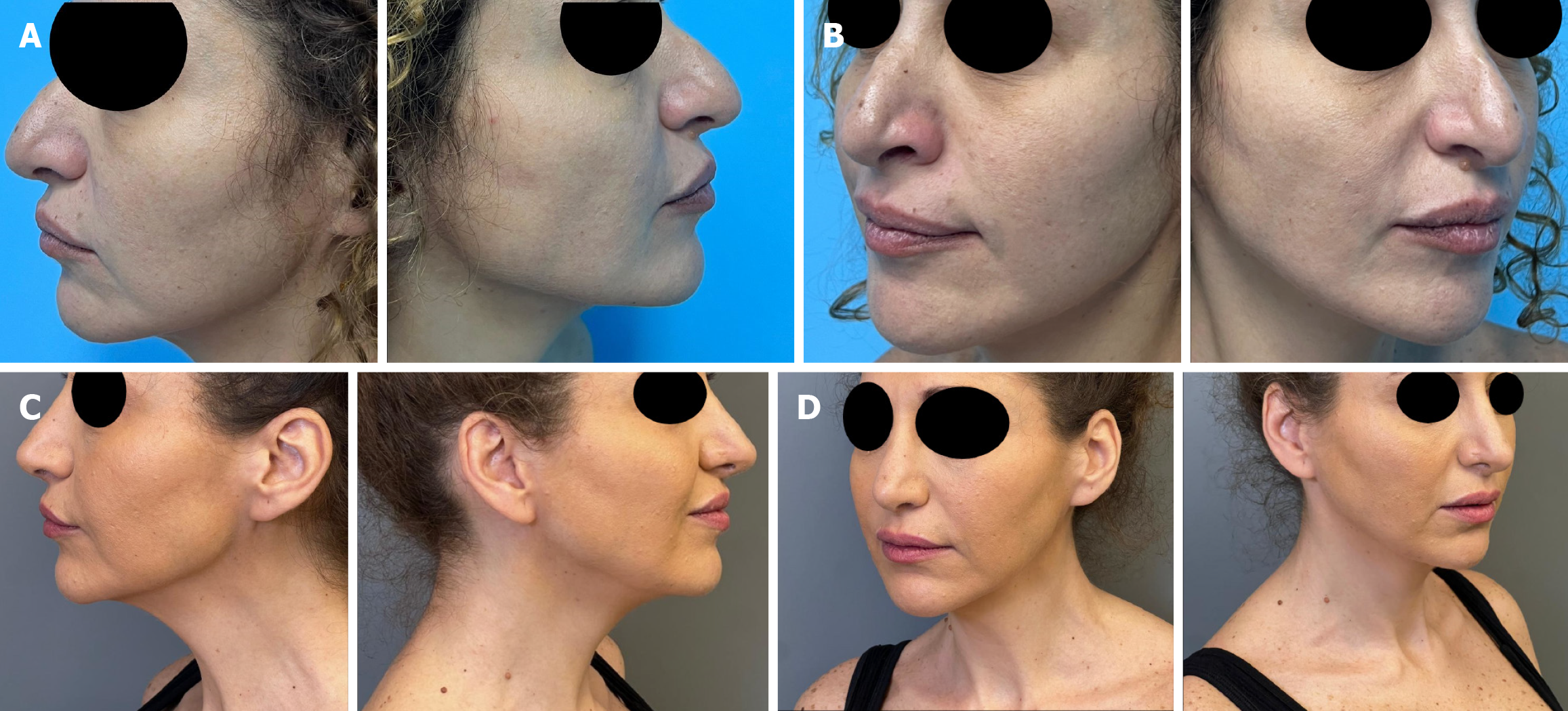
She presented for a revision rhinoplasty to address nasal deformities, including pollybeak deformity, a droopy tip with alar notching, and an over-resected bony dorsum (Figure 1). She was also dissatisfied with the results of her breast surgery due to lateral displacement of the implants and bilateral breast bottoming out.
Prior to presentation, the patient had undergone rhinoplasty elsewhere 10 years ago and an augmentation mastopexy with retropectoral 275cc full-profile nanotextured Motiva implants 3 years ago.
Given these concerns, we decided to combine the secondary rhinoplasty with the breast revision in the same surgery under general anesthesia.
During the breast revision surgery, a 4 cm × 4 cm fragment of the periprosthetic capsule was harvested, along with a rib graft from the right 5th rib. A novel technique called DCIC was employed for nasal dorsum augmentation. This technique aims to augment the nasal dorsum while minimizing the risk of graft warping or visibility.
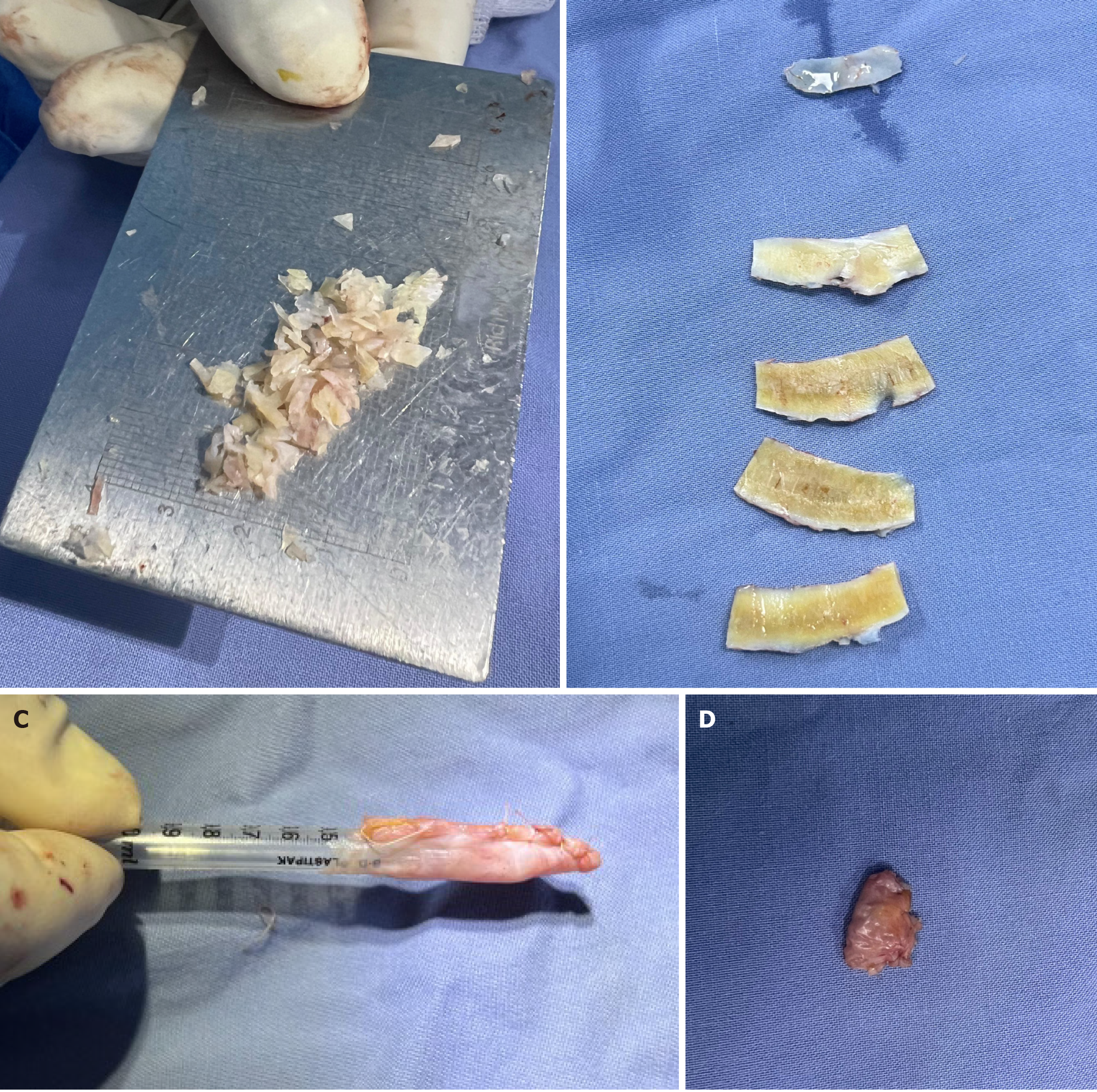
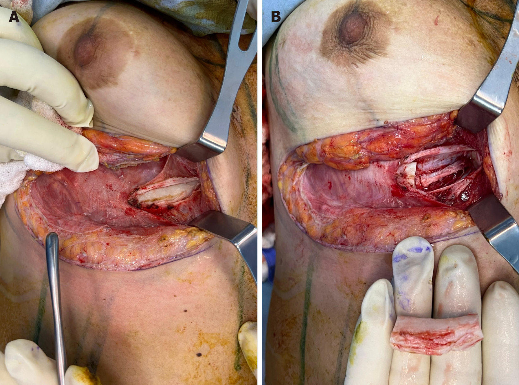
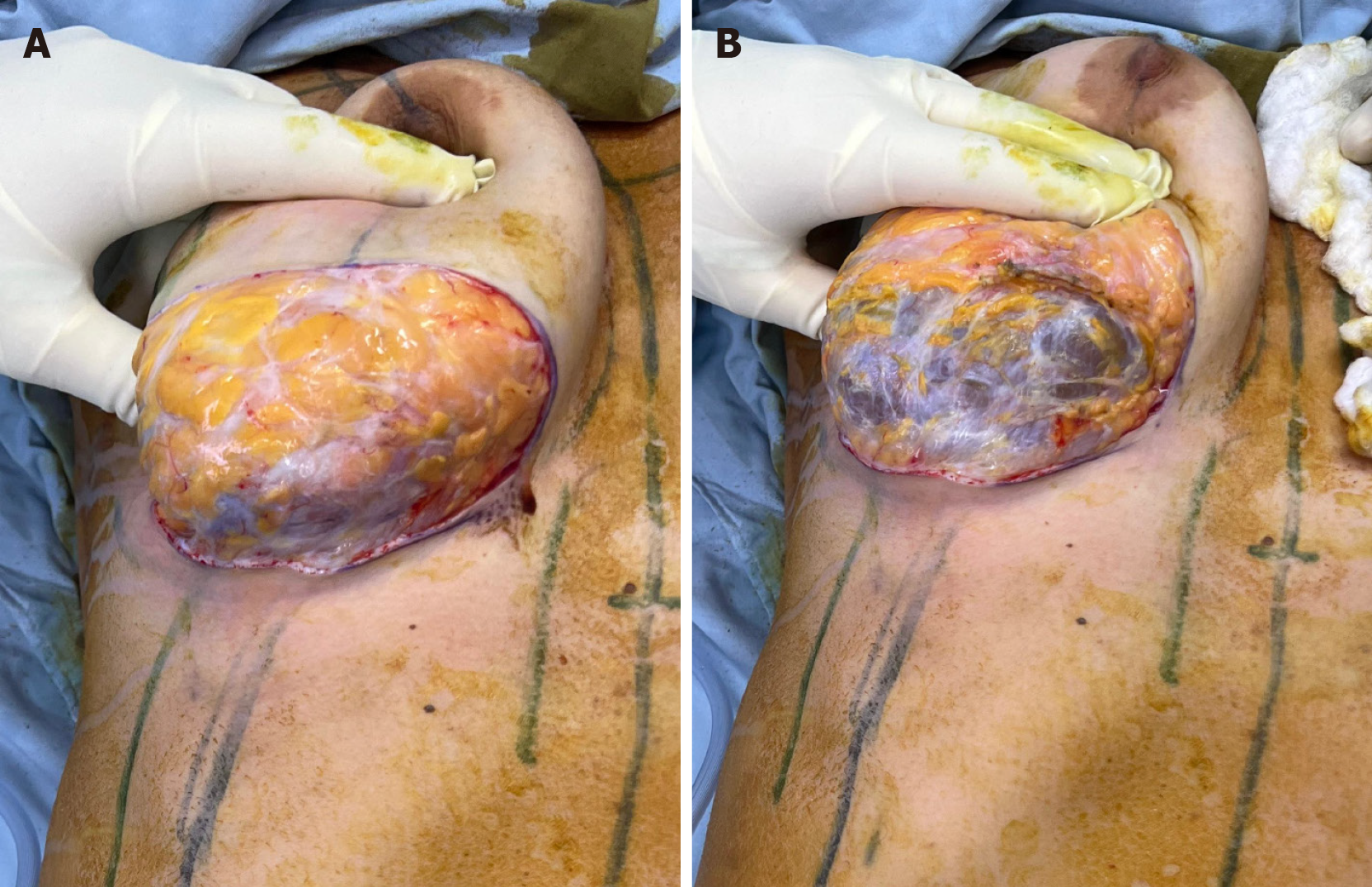
The surgical procedure began with a revision mastopexy with implant exchange. A right submammary fold incision was made, and the periprosthetic capsule was identified. The intact implant was removed. A 4 cm × 4 cm fragment of the capsule was harvested and defatted. The cartilage of the right 5th rib was identified and meticulously dissected in a subperichondrial plane (Figure 2A). The entire rib cartilage was harvested (Figures 3 and 4). A Valsalva maneuver was performed to confirm the absence of a pleural injury. The perichondrium was approximated using Vicryl 2/0 simple sutures, and the pectoralis muscle aponeurosis was sutured. The retropectoral implant pocket was reduced laterally on both breasts, and new Mentor textured (Siltex) 325cc Xtragel high-profile implants were inserted. An inverted-T mastopexy was then performed.
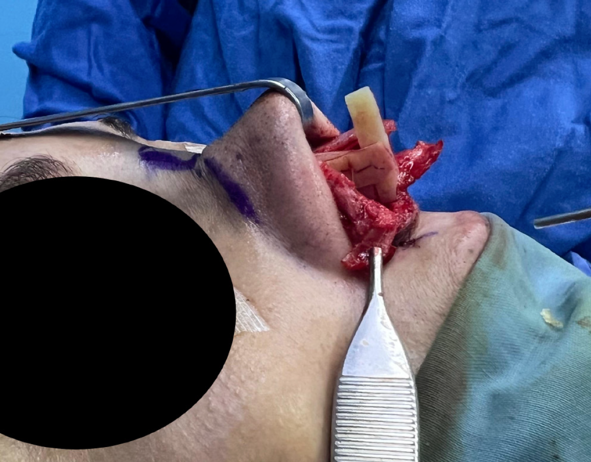
The rhinoplasty began with reopening the previous incision, followed by meticulous dissection of the nasal structures. Supratip fibrosis was excised, excess upper lateral cartilage was trimmed, and anterior septal resection was performed to correct the pollybeak deformity. A septal cartilage graft was harvested, ensuring a residual 15 mm L-strut was preserved for structural integrity. The harvested rib cartilage was then carefully sculpted (Figure 2B) into multiple lamellae. Bilateral extended spreader grafts were sutured in place. A columellar strut was introduced between the spreader grafts and sutured to them (Figure 5), providing strong tip support and preventing future tip deviation.
The intermediate crura of the alar cartilages were found to be interrupted. The medial crura were shortened to decrease tip projection and were sutured to the columellar strut. Bilateral underlay septal cartilage grafts were placed to strengthen the lateral crura. The medial crura were sutured to the lateral crura bilaterally. A dome equalizing suture with a 5/0 Prolene suture was performed. This tip modification strengthens the tip, decreases projection, and enhances rotation, all while being covered with thick fibrotic skin.
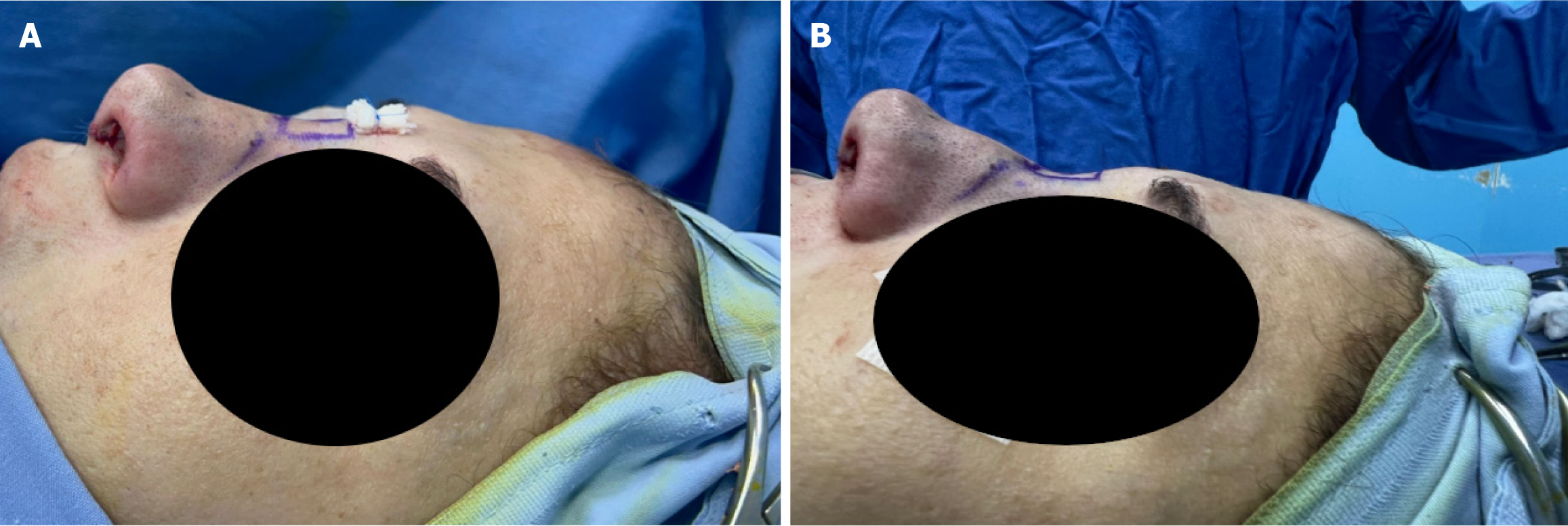
The capsular fragment was sutured together over a 1 cc syringe in a tube-like fashion (Figure 2C) using Vicryl Rapide 4/0. The remaining rib cartilage was diced into very small fragments (Figure 2A) and introduced into the capsular tube, which was then sealed with Vicryl 4/0 Rapide sutures (Figure 2D). The DCIC graft was inserted over the nasal bones and stabilized with a transcutaneous Prolene 5/0 suture over a tulle gras gauze (Figure 6A). This technique provides dorsal augmentation and straightens the nasal dorsum (Figure 6).
Bilateral alar contour grafts were placed to reduce alar rim notching. The columellar skin was closed using nylon 6/0, and the mucosa was sutured with Vicryl Rapide 5/0. Packing remained in place for 48 hours, and the nasal splint, columellar sutures, and Prolene suture were removed after eight days.
At 12 months follow-up, the patient has tolerated the surgery very well, showing no signs of complications in the nasal or chest region. There were no breathing difficulties, nasal irregularities, or chest-related symptoms, further reinforcing the suitability of the DCIC technique.
We present a case of the DCIC technique, in which the DCIF technique was modified by using periprosthetic capsule material from a previous breast augmentation mastopexy instead of fascia from the rectus abdominis or temporalis[8]. This patient, who had undergone breast augmentation mastopexy three years prior, required revisional surgery due to lateral displacement of the implants and breast asymmetry. This circumstance provided an opportunity to use the capsular material as a novel graft wrapper[9].
The use of alternative materials, such as deep temporalis fascia and rectus abdominis aponeurosis, has been explored in other rhinoplasty techniques. These materials serve similar purposes in providing a structural and aesthetic framework for nasal grafts. However, the innovative use of breast capsular material in this context not only repurposes biological waste but also reduces scarring by minimizing the need for additional donor site harvesting.
Using this autologous material, which is naturally biocompatible, offers a unique approach to enhancing graft integration and minimizing foreign body reactions.
At the 12-month follow-up, the patient had tolerated the surgery very well, showing no signs of complications in the nasal or chest regions. There were no breathing difficulties, nasal irregularities, or chest-related symptoms, further reinforcing the suitability of the DCIC technique. Additionally, a comparison between DCIC and traditional methods (DCIF and Turkish Delight) showed a comparable healing time to DCIF but faster healing than the traditional Turkish Delight method, likely due to the biocompatibility of the periprosthetic capsule. The reduced warping, visibility, and palpability in DCIC compared to DCIF suggests superior patient outcomes in dorsum contouring and structural support. The well-vascularized nature of this tissue might enhance graft integration, promoting quicker healing and better overall outcomes. Using DCIC eliminates the risk of graft warping, provides a smoother dorsum contour, minimizes visibility and palpability, and eliminates the need for additional donor site harvesting. The capsule's biocompatibility and the absence of reactive inner tissue further minimize the risk of complications, such as inflammation and infection.
However, it is important to note that the application of this technique is limited to specific cases, particularly those involving patients undergoing concurrent revision breast implant surgery and rhinoplasty, as demonstrated in our case. The DCIC technique adds a new tool to our surgical armamentarium, applicable in selected cases.
Potential complications include graft rejection, though this risk remains minimal due to the autologous nature of the capsule. Infection rates appear to be lower compared to synthetic materials, and material resorption is limited due to the stability of encapsulated cartilage. Fibrosis is comparable to fascia-based techniques but without additional donor site morbidity.
However, it is essential to recognize that this study presents a single case report of an innovative technique. Larger, prospective studies comparing DCIC to traditional methods are needed to establish its efficacy and potentially refine and develop the technique further.
This case highlights the innovative adaptation of the Turkish Delight technique through the DCIC method, which utilizes periprosthetic capsular material. Our findings suggest that using autologous materials, such as the breast capsule, offers significant advantages in biocompatibility and graft integration, with no complications observed at six months postoperatively. A comprehensive meta-analysis indicates that the most common long-term complications of diced cartilage grafts in rhinoplasty include infection (4.5%) and visible irregularity (5.3%), with a 5.3% revision surgery rate[10]. The DCIC technique, which utilizes readily available autologous material, could potentially reduce these complication rates and improve long-term outcomes in rhinoplasty. This adaptation underscores the importance of innovation in surgical practices, particularly in cosmetic surgery, where patient-specific characteristics and previous surgical histories can be transformed into opportunities for improved outcomes.
| 1. | Moretti A, Sciuto S. Rib grafts in septorhinoplasty. Acta Otorhinolaryngol Ital. 2013;33:190-195. [PubMed] |
| 2. | Cakmak O, Ergin T. The versatile autogenous costal cartilage graft in septorhinoplasty. Arch Facial Plast Surg. 2002;4:172-176. [RCA] [PubMed] [DOI] [Full Text] [Cited by in Crossref: 41] [Cited by in RCA: 43] [Article Influence: 1.8] [Reference Citation Analysis (0)] |
| 3. | Moshaver A, Gantous A. The use of autogenous costal cartilage graft in septorhinoplasty. Otolaryngol Head Neck Surg. 2007;137:862-867. [RCA] [PubMed] [DOI] [Full Text] [Cited by in Crossref: 33] [Cited by in RCA: 38] [Article Influence: 2.1] [Reference Citation Analysis (0)] |
| 4. | Erol OO. The Turkish delight: a pliable graft for rhinoplasty. Plast Reconstr Surg. 2000;105:2229-41; discussion 2242. [RCA] [PubMed] [DOI] [Full Text] [Cited by in Crossref: 234] [Cited by in RCA: 246] [Article Influence: 9.5] [Reference Citation Analysis (0)] |
| 5. | Wright JM, Halsey JN, Rottgers SA. Dorsal Augmentation: A Review of Current Graft Options. Eplasty. 2023;23:e4. [PubMed] |
| 6. | Yoo SH, Jang YJ. Rib cartilage in Asian rhinoplasty: new trends. Curr Opin Otolaryngol Head Neck Surg. 2019;27:261-266. [RCA] [PubMed] [DOI] [Full Text] [Cited by in Crossref: 21] [Cited by in RCA: 42] [Article Influence: 7.0] [Reference Citation Analysis (0)] |
| 7. | Dresner HS, Hilger PA. An overview of nasal dorsal augmentation. Semin Plast Surg. 2008;22:65-73. [RCA] [PubMed] [DOI] [Full Text] [Cited by in Crossref: 27] [Cited by in RCA: 34] [Article Influence: 2.3] [Reference Citation Analysis (0)] |
| 8. | As'adi K, Salehi SH, Shoar S. Rib Diced Cartilage-Fascia Grafting in Dorsal Nasal Reconstruction: A Randomized Clinical Trial of Wrapping With Rectus Muscle Fascia vs Deep Temporal Fascia. Aesthet Surg J. 2014;34:NP21-NP31. [RCA] [PubMed] [DOI] [Full Text] [Cited by in Crossref: 14] [Cited by in RCA: 19] [Article Influence: 1.6] [Reference Citation Analysis (0)] |
| 9. | Rohrich RJ, Agrawal N, Avashia Y, Savetsky IL. Safety in the Use of Fillers in Nasal Augmentation-the Liquid Rhinoplasty. Plast Reconstr Surg Glob Open. 2020;8:e2820. [RCA] [PubMed] [DOI] [Full Text] [Full Text (PDF)] [Cited by in Crossref: 1] [Cited by in RCA: 7] [Article Influence: 1.2] [Reference Citation Analysis (0)] |
| 10. | Li J, Sang C, Fu R, Liu C, Suo L, Yan Y, Liu K, Huang RL. Long-Term Complications from Diced Cartilage in Rhinoplasty: A Meta-Analysis. Facial Plast Surg Aesthet Med. 2022;24:221-227. [RCA] [PubMed] [DOI] [Full Text] [Cited by in Crossref: 2] [Cited by in RCA: 13] [Article Influence: 2.6] [Reference Citation Analysis (0)] |


