Editorial Open Access
Copyright ©The Author(s) 2024. Published by Baishideng Publishing Group Inc. All rights reserved.
World J Clin Cases. Oct 26, 2024; 12(30): 6353-6357
Published online Oct 26, 2024. doi: 10.12998/wjcc.v12.i30.6353
Global strategy for prevention of gastric cancer
Sergey M Kotelevets, Department of Therapy, North Caucasus State Academy, Cherkessk 369000, Russia
ORCID number: Sergey M Kotelevets (0000-0003-4915-6869).
Author contributions: Kotelevets SM wrote the entire manuscript, and read and approved the final manuscript.
Conflict-of-interest statement: The author declare that they have no potential conflicts of interest to disclose.
Open-Access: This article is an open-access article that was selected by an in-house editor and fully peer-reviewed by external reviewers. It is distributed in accordance with the Creative Commons Attribution NonCommercial (CC BY-NC 4.0) license, which permits others to distribute, remix, adapt, build upon this work non-commercially, and license their derivative works on different terms, provided the original work is properly cited and the use is non-commercial. See: https://creativecommons.org/Licenses/by-nc/4.0/
Corresponding author: Sergey M Kotelevets, MD, Associate Professor, Department of Therapy, North Caucasus State Academy, Lenina Street 75/32, Cherkessk 369000, Russia. smkotelevets@mail.ru
Received: June 15, 2024
Revised: July 9, 2024
Accepted: July 15, 2024
Published online: October 26, 2024
Processing time: 80 Days and 20.3 Hours

Abstract

Global prevention of gastric cancer needs to increase its level of effectiveness. The prevention strategy should include all stages of primary and secondary prevention. The necessary steps to prevent gastric cancer are the following: Maintaining a healthy lifestyle and diet, avoiding smoking and alcohol; serological screening of Helicobacter pylori infections and eradication; serological screening of atrophic gastritis in the population over 45 years of age and identification of severe atrophic gastritis with a high risk of developing gastric cancer; verification of atrophic gastritis and precancerous changes in the gastric mucosa using modern endoscopic (confocal laser endomicroscopy, narrow-spectrum imaging, and magnifying endoscopy) and morphological methods among patients with severe atrophic gastritis who were identified using serological screening; treatment of patients with atrophic gastritis during diagnosis verification; annual endoscopic and morphological monitoring of patients with atrophic gastritis during permanent treatment; annual serological monitoring of patients with atrophic gastritis who refused endoscopic and morphological monitoring; and radical treatment of patients with verified early gastric cancer. Ways to implement the algorithm for the global strategy for the prevention of gastric cancer (protocol of practical recommendations) are: State, government, and municipal programs; departmental programs of health departments; family doctors for patients who have a contract at the initiative of the doctor; family doctors for patients with a contract at the patient’s initiative; and within private healthcare system where both doctors and patients can initiate the implementation of algorithm.

Key Words: Prevention; Gastric cancer; Strategy; Atrophic gastritis; Serological screening; Precancerous changes

Core Tip: Many different models of primary and secondary prevention of gastric cancer have been proposed at present. However, there is no global consistent strategy and protocol for practical recommendations. The lack of a specific concept does not effectively reduce the incidence and mortality of gastric cancer. In this article, the author proposes a global preventive strategy for the prevention of gastric cancer and ways to implement it. The article also contains an algorithm of actions that takes into account the individual conditions of its use.


INTRODUCTION

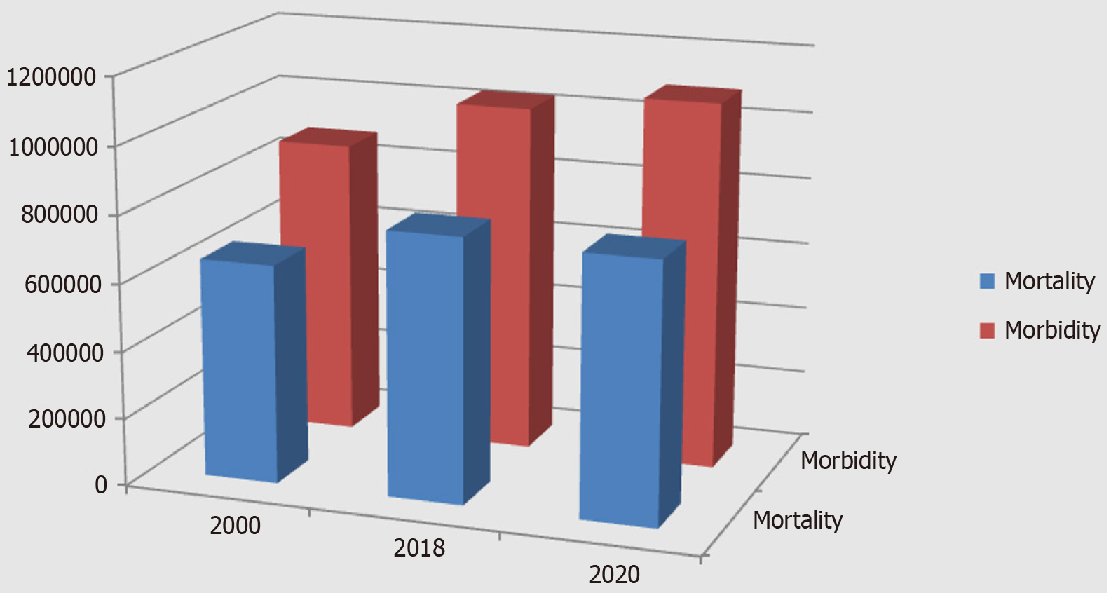
In 2000, 880000 people worldwide were diagnosed with stomach cancer and 650000 died from it[1]. Since then, there has been a steady increase in global statistical indicators of morbidity and mortality for stomach cancer. For example, in 2018, stomach cancer was the second leading cause of cancer-related death and the sixth most common cancer in the world. A total of 1033701 new cases of stomach cancer were reported in 2018, and 782685 patients have died from the disease (Figure 1). This number of deaths from stomach cancer accounts for about 8.2% of global cancer deaths[2]. In 2020, 1089103 people developed stomach cancer in the world, and 768793 of them died[3]. The disease still holds a leading position. Fifty percent of patients with stomach cancer and deaths from stomach cancer are residents of China[4]. The prognosis for the next few years is negative, and a further increase in the incidence of stomach cancer is expected[5]. The difference between the indicators of global population growth, the incidence of stomach cancer, and the death rate from stomach cancer is significant (P = 0.000). Therefore, it is necessary to distinguish between absolute and relative indicators of the 20-year dynamics of the incidence of stomach cancer and mortality from the disease. For 20 years, world statistics on stomach cancer have been characterized by an increase in such indicators as morbidity and mortality (in absolute terms). At the same time, the world's population has been characterized by an even more pronounced growth over the same period. Consequently, relative to the global population, there is a decrease in the incidence of stomach cancer and a more significant decrease in mortality from stomach cancer. Since 2020, new screening technologies for the detection, diagnosis, and treatment of precancerous conditions of the stomach have appeared and achieved perfection. First of all, this is the appearance of highly sensitive and highly specific serological markers for gastric precancerous conditions (atrophic gastritis), which allow effective screening of precancerous gastric pathology in the population of any number belonging to the risk group (40 +). Narrow-spectrum and magnifying endoscopy, using modern endoscopic equipment such as Olympus EXERA III GIF-190 HQ, Olympus EVIS X1, has achieved incredible success in the diagnosis of precancerous diseases and precancerous changes in the gastric mucosa. Narrowband imaging, texture and color enhancement imaging, red dichromatic imaging, and other image-enhanced technologies (image-enhanced endoscopy) have become frequently used in practical healthcare. In addition, the active introduction of computer detection using artificial intelligence in endoscopy has begun. An increase in magnification of 540 times has become available during endoscopic examination of the gastric mucosa (confocal laser endomicroscopy of the upper digestive tract allows a magnification to be increased to a thousand times). As a result, the diagnostic capabilities for detecting precancerous changes in the gastric mucosa have significantly increased[6].

Figure 1
Figure 1 Dynamics of morbidity and mortality from 2000 to 2020 (statistics on stomach cancer).

The dynamics of global statistics on morbidity and mortality due to gastric cancer show unsatisfactory results (Figure 1): (1) There is a decrease in the incidence and mortality of stomach cancer, relative to the growth of the world's population, from 2000 to 2020; (2) The positive dynamics of the incidence and mortality of stomach cancer is very poor, relative to the growth of the world's population (morbidity decreased by 3.5% and mortality by 9%) for 20 years; and (3) This indicates the low effectiveness of preventive and therapeutic measures for stomach cancer, which is associated with the insufficient implementation of modern screening and diagnostic technologies.

There are substantial, high-quality data supporting the effective prevention of gastric cancer that are derived from international studies[7]. Health systems use traditional primary and secondary prevention methods for gastric cancer. Primary prevention of gastric cancer includes mass screening and treatment programs for Helicobacter pylori (H. pylori) infection (“test and treat”), reducing consumption of tobacco and alcohol, and obesity control[8-11]. However, the role of other factors, such as lifestyle, diet, and drug use is still under debate in gastric carcinogenesis[12]. Secondary prevention of gastric cancer includes the diagnosis of early gastric cancer using narrow-band endoscopic imaging methods[13,14]. Some authors suggest introducing screening for atrophic gastritis and precancerous changes in the gastric mucosa to determine the level of risk of developing gastric cancer[15]. What type of prevention is this, primary or secondary? Researchers in some countries propose combining primary and secondary prevention methods to increase the effectiveness of gastric cancer prevention[15,16]. The financial burden makes it very difficult to implement various preventive actions in the population[17,18,19]. A consensus protocol for use in gastric cancer prevention, the global gastric cancer prevention strategy, is currently needed because traditional primary and secondary prevention methods are not effective.

CLASSIFICATION AND FEATURES OF GASTRIC CANCER

Lauren's classification defines two types of gastric cancer: Intestinal-type and diffuse-type[20]. The role of H. pylori in the carcinogenesis of gastric cancer of intestinal and diffuse types has been confirmed by many authors[21,22]. A comparative study of the entire complex of clinical, morphological, prognostic, and immunohistochemical characteristics and parenchymal-stromal relationships in gastric cancer allows us to confirm the feasibility of identifying three main histological classification forms of gastric cancer: Intestinal, diffuse, and mixed. The additional identification of the mixed form of gastric cancer includes significant, early unclassified tumors of complex structure with multidirectional differentiation of the cancerous epithelium according to Correa's cascade type[23,24].

STEPS OF PROTOCOL FOR GLOBAL STRATEGY FOR PREVENTION OF GASTRIC CANCER

The absence of symptoms is an important characteristic of early gastric cancer and precancerous diseases and changes. Such patients are not examined by modern endoscopic methods, such as confocal laser endomicroscopy, narrow-band imaging, and magnifying endoscopy. Modern endoscopic methods are not used for primary screening of early gastric cancer and precancerous lesions due to the high cost of equipment and sensors, as well as the need for trained specialists to interpret images. Invasive endoscopic methods can only be used to verify the diagnosis of precancerous changes and early gastric cancer in patients identified by screening methods. The population has no motivation for examination using invasive endoscopic methods in the absence of symptoms. The identified precancerous pathology (atrophic gastritis) by serological screening is the basis for motivating the patient to continue examination using endoscopic methods with biopsy. Morphological verification of precancerous pathology with a high risk of developing gastric cancer motivates the patient for annual endoscopic monitoring. Morphological examination of biopsy specimens is the “gold” standard for diagnosis, despite the high level of resources of modern endoscopic technologies. Morphological examination of biopsy specimens may not always be the reference standard for diagnosis: Example 1: Morphological diagnosis of the focus of gastric cancer is the reference standard for diagnosis; Example 2: Morphological diagnosis of atrophic gastritis is the reference standard for diagnosis; and Example 3: Morphological examination of biopsy samples of the gastric mucosa is not the standard for determining the level of risk of developing gastric cancer. The risk of developing gastric cancer is proportional to the severity of mucosal atrophy, with both ranging from low to high[25]. Atrophic gastritis is a multifocal atrophic process in the gastric mucosa. The gastric mucosa of different localizations has different degrees of severity of mucosal atrophy. How do you determine the integral severity of mucosal atrophy on the total surface of the stomach? The total surface area of the stomach ranges from 220 ± 27 cm2 to 536 ± 25 cm2. Changes in total gastric volume and total gastric surface area are associated with food intake and gastric emptying. During endoscopy, this is associated with air insufflation[26]. The severity of atrophy of the gastric mucosa is identified in the biopsy specimen in accordance with the morphological visual analogue scale and is determined by the degree of reduction in the number of gastric glands. The area of each biopsy is several mm2. It is impossible to calculate how much surface area of the stomach has mild, moderate, or severe atrophy using a biopsy. Using endoscopic visualization, it is possible to determine the surface area of the stomach with atrophy in accordance with the Kimura-Takemoto classification from C1 to O3. But modern endoscopic methods cannot reveal the severity of atrophy of the gastric mucosa in a specific surface area of the stomach. Endoscopic morphometry is not possible. It is unrealistic to assess the depth (severity) of atrophy of the gastric mucosa using modern endoscopic methods. The risk of developing gastric cancer cannot, therefore, be precisely detailed. The updated Kimura-Takemoto classification of atrophic gastritis is the most optimal way to assess the severity of atrophic gastritis and the risk of developing gastric cancer[27].

CONCLUSION

Global prevention of gastric cancer needs to increase its level of effectiveness. The prevention strategy should include all stages of primary and secondary prevention. The necessary steps to prevent gastric cancer are the following: (1) Maintaining a healthy lifestyle and diet, avoiding smoking and alcohol; (2) Serological screening of H. pylori infections and eradication; (3) Serological screening of atrophic gastritis in the population over 45 years of age and identification of severe atrophic gastritis with a high risk of developing gastric cancer; (4) Verification of atrophic gastritis and precancerous changes in the gastric mucosa using modern endoscopic (confocal laser endomicroscopy, narrow-spectrum imaging, and magnifying endoscopy) and morphological methods among patients with severe atrophic gastritis who were identified using serological screening; (5) Treatment of patients with atrophic gastritis during diagnosis verification; (6) Annual endoscopic and morphological monitoring of patients with atrophic gastritis during permanent treatment; (7) Annual serological monitoring of patients with atrophic gastritis who refused endoscopic and morphological monitoring; and (8) Radical treatment of patients with verified early gastric cancer.

Ways to implement the algorithm for the global strategy for the prevention of gastric cancer (protocol of practical recommendations): (1) State, government, and municipal programs; (2) Departmental programs of health departments; (3) Family doctors for patients who have a contract at the initiative of the doctor; (4) Family doctors for patients with a contract at the patient’s initiative; and (5) Within private healthcare system where both doctors and patients can initiate the implementation of algorithm.

Footnotes

Provenance and peer review: Invited article; Externally peer reviewed.

Peer-review model: Single blind

Specialty type: Medicine, research and experimental

Country of origin: Russia

Peer-review report’s classification

Scientific Quality: Grade C

Novelty: Grade B

Creativity or Innovation: Grade B

Scientific Significance: Grade B

P-Reviewer: Amin KFM S-Editor: Luo ML L-Editor: Wang TQ P-Editor: Zheng XM

References
1.  Crew KD, Neugut AI. Epidemiology of gastric cancer. World J Gastroenterol. 2006;12:354-362.  [PubMed]  [DOI]  [Cited in This Article: ]  [Cited by in CrossRef: 1193]  [Cited by in F6Publishing: 1220]  [Article Influence: 67.8]  [Reference Citation Analysis (8)]
2.  Bray F, Ferlay J, Soerjomataram I, Siegel RL, Torre LA, Jemal A. Global cancer statistics 2018: GLOBOCAN estimates of incidence and mortality worldwide for 36 cancers in 185 countries. CA Cancer J Clin. 2018;68:394-424.  [PubMed]  [DOI]  [Cited in This Article: ]  [Cited by in Crossref: 53206]  [Cited by in F6Publishing: 53419]  [Article Influence: 8903.2]  [Reference Citation Analysis (124)]
3.  Sung H, Ferlay J, Siegel RL, Laversanne M, Soerjomataram I, Jemal A, Bray F. Global Cancer Statistics 2020: GLOBOCAN Estimates of Incidence and Mortality Worldwide for 36 Cancers in 185 Countries. CA Cancer J Clin. 2021;71:209-249.  [PubMed]  [DOI]  [Cited in This Article: ]  [Cited by in Crossref: 50630]  [Cited by in F6Publishing: 54562]  [Article Influence: 18187.3]  [Reference Citation Analysis (156)]
4.  Kang B, Liu XY, Cheng YX, Tao W, Peng D. Factors associated with hypertension remission after gastrectomy for gastric cancer patients. World J Gastrointest Surg. 2022;14:743-753.  [PubMed]  [DOI]  [Cited in This Article: ]  [Cited by in F6Publishing: 4]  [Reference Citation Analysis (0)]
5.  Smyth EC, Nilsson M, Grabsch HI, van Grieken NC, Lordick F. Gastric cancer. Lancet. 2020;396:635-648.  [PubMed]  [DOI]  [Cited in This Article: ]  [Cited by in Crossref: 1150]  [Cited by in F6Publishing: 2270]  [Article Influence: 567.5]  [Reference Citation Analysis (0)]
6.  Bayguinov PO, Oakley DM, Shih CC, Geanon DJ, Joens MS, Fitzpatrick JAJ. Modern Laser Scanning Confocal Microscopy. Curr Protoc Cytom. 2018;85:e39.  [PubMed]  [DOI]  [Cited in This Article: ]  [Cited by in Crossref: 48]  [Cited by in F6Publishing: 49]  [Article Influence: 8.2]  [Reference Citation Analysis (0)]
7.  Huang RJ, Epplein M, Hamashima C, Choi IJ, Lee E, Deapen D, Woo Y, Tran T, Shah SC, Inadomi JM, Greenwald DA, Hwang JH. An Approach to the Primary and Secondary Prevention of Gastric Cancer in the United States. Clin Gastroenterol Hepatol. 2022;20:2218-2228.e2.  [PubMed]  [DOI]  [Cited in This Article: ]  [Cited by in Crossref: 11]  [Cited by in F6Publishing: 18]  [Article Influence: 9.0]  [Reference Citation Analysis (0)]
8.  Corsi Sotelo Ó, Pizarro Rojas M, Rollán Rodríguez A, Silva Figueroa V, Araya Jofré R, Bufadel Godoy ME, Cortés González P, González Donoso R, Fuentes López E, Latorre Selvat G, Medel-Jara P, Reyes Placencia D, Pizarro Véliz M, Garchitorena Marqués MJ, Zegers Vial MT, Crispi Galleguillos F, Espinoza MA, Riquelme Pérez A. Chilean consensus by expert panel using the Delphi technique for primary and secondary prevention of gastric cancer. Gastroenterol Hepatol. 2024;S0210-5705(24)00026.  [PubMed]  [DOI]  [Cited in This Article: ]  [Cited by in Crossref: 2]  [Reference Citation Analysis (0)]
9.  Huang RJ, Laszkowska M, In H, Hwang JH, Epplein M. Controlling Gastric Cancer in a World of Heterogeneous Risk. Gastroenterology. 2023;164:736-751.  [PubMed]  [DOI]  [Cited in This Article: ]  [Cited by in Crossref: 20]  [Cited by in F6Publishing: 27]  [Article Influence: 27.0]  [Reference Citation Analysis (0)]
10.  Arnold M, Abnet CC, Neale RE, Vignat J, Giovannucci EL, McGlynn KA, Bray F. Global Burden of 5 Major Types of Gastrointestinal Cancer. Gastroenterology. 2020;159:335-349.e15.  [PubMed]  [DOI]  [Cited in This Article: ]  [Cited by in Crossref: 857]  [Cited by in F6Publishing: 971]  [Article Influence: 242.8]  [Reference Citation Analysis (0)]
11.  Cheng XJ, Lin JC, Tu SP. Etiology and Prevention of Gastric Cancer. Gastrointest Tumors. 2016;3:25-36.  [PubMed]  [DOI]  [Cited in This Article: ]  [Cited by in Crossref: 97]  [Cited by in F6Publishing: 98]  [Article Influence: 12.3]  [Reference Citation Analysis (0)]
12.  Eusebi LH, Telese A, Marasco G, Bazzoli F, Zagari RM. Gastric cancer prevention strategies: A global perspective. J Gastroenterol Hepatol. 2020;35:1495-1502.  [PubMed]  [DOI]  [Cited in This Article: ]  [Cited by in Crossref: 93]  [Cited by in F6Publishing: 148]  [Article Influence: 37.0]  [Reference Citation Analysis (0)]
13.  Yoshida N, Doyama H, Yano T, Horimatsu T, Uedo N, Yamamoto Y, Kakushima N, Kanzaki H, Hori S, Yao K, Oda I, Katada C, Yokoi C, Ohata K, Yoshimura K, Ishikawa H, Muto M. Early gastric cancer detection in high-risk patients: a multicentre randomised controlled trial on the effect of second-generation narrow band imaging. Gut. 2021;70:67-75.  [PubMed]  [DOI]  [Cited in This Article: ]  [Cited by in Crossref: 50]  [Cited by in F6Publishing: 71]  [Article Influence: 23.7]  [Reference Citation Analysis (0)]
14.  Kim YI, Choi IJ. Current Evidence for a Paradigm Shift in Gastric Cancer Prevention From Endoscopic Screening to Helicobacter pylori Eradication in Korea. J Gastric Cancer. 2022;22:169-183.  [PubMed]  [DOI]  [Cited in This Article: ]  [Reference Citation Analysis (0)]
15.  Farinati F, Pelizzaro F. Gastric cancer screening in Western countries: A call to action. Dig Liver Dis. 2024;24:S1590-8658(24)00269.  [PubMed]  [DOI]  [Cited in This Article: ]  [Cited by in Crossref: 2]  [Reference Citation Analysis (0)]
16.  Rugge M, Sugano K, Scarpignato C, Sacchi D, Oblitas WJ, Naccarato AG. Gastric cancer prevention targeted on risk assessment: Gastritis OLGA staging. Helicobacter. 2019;24:e12571.  [PubMed]  [DOI]  [Cited in This Article: ]  [Cited by in Crossref: 18]  [Cited by in F6Publishing: 16]  [Article Influence: 3.2]  [Reference Citation Analysis (0)]
17.  Hu Z, Liu Z, Li W, You W, Pan K. Health economic evaluation on population-based Helicobacter pylori eradication and endoscopic screening for gastric cancer prevention. Chin J Cancer Res. 2023;35:595-605.  [PubMed]  [DOI]  [Cited in This Article: ]  [Reference Citation Analysis (0)]
18.  Kowada A. A Population-Based Helicobacter pylori Eradication Strategy Is More Cost-Effective than Endoscopic Screening. Dig Dis Sci. 2023;68:1735-1746.  [PubMed]  [DOI]  [Cited in This Article: ]  [Cited by in F6Publishing: 11]  [Reference Citation Analysis (0)]
19.  Gotoda T, Ishikawa H, Kusano C, Suzuki S, Ohnishi H, Sugano K, Matsuyama Y. Randomized controlled trial comparing the costs of gastric cancer screening systems between serological risk-based upper gastrointestinal endoscopy and the existing barium photofluorography: gastric cancer screening labeled by serum examination in place of aged gastric cancer organized screening systems (GALAPAGOS study). Gastric Cancer. 2024;27:36-48.  [PubMed]  [DOI]  [Cited in This Article: ]  [Cited by in Crossref: 1]  [Reference Citation Analysis (0)]
20.  Lauren P. The Two Histological Main Types of Gastric Carcinoma: Diffuse and So-Called Intestinal-Type Carcinoma. An Attempt at A Histo-Clinical Classification. Acta Pathol Microbiol Scand. 1965;64:31-49.  [PubMed]  [DOI]  [Cited in This Article: ]  [Cited by in Crossref: 4011]  [Cited by in F6Publishing: 4203]  [Article Influence: 150.1]  [Reference Citation Analysis (0)]
21.  Ansari S, Gantuya B, Tuan VP, Yamaoka Y. Diffuse Gastric Cancer: A Summary of Analogous Contributing Factors for Its Molecular Pathogenicity. Int J Mol Sci. 2018;19:2424.  [PubMed]  [DOI]  [Cited in This Article: ]  [Cited by in Crossref: 40]  [Cited by in F6Publishing: 55]  [Article Influence: 9.2]  [Reference Citation Analysis (0)]
22.  Moss SF. The Clinical Evidence Linking Helicobacter pylori to Gastric Cancer. Cell Mol Gastroenterol Hepatol. 2017;3:183-191.  [PubMed]  [DOI]  [Cited in This Article: ]  [Cited by in Crossref: 149]  [Cited by in F6Publishing: 186]  [Article Influence: 23.3]  [Reference Citation Analysis (0)]
23.  Danilova IA, Anichkov NM. [Comprehensive study of the major morphological forms of gastric cancer in relation to the indices of their prognostic assessment]. Arkh Patol. 2009;71:27-31.  [PubMed]  [DOI]  [Cited in This Article: ]
24.  Fang X, Wu X, Xiang E, Luo F, Li Q, Ma Q, Yuan F, Chen P. Expression profiling of CPS1 in Correa's cascade and its association with gastric cancer prognosis. Oncol Lett. 2021;21:441.  [PubMed]  [DOI]  [Cited in This Article: ]  [Cited by in Crossref: 2]  [Cited by in F6Publishing: 2]  [Article Influence: 0.7]  [Reference Citation Analysis (0)]
25.  Sugano K, Tack J, Kuipers EJ, Graham DY, El-Omar EM, Miura S, Haruma K, Asaka M, Uemura N, Malfertheiner P; faculty members of Kyoto Global Consensus Conference. Kyoto global consensus report on Helicobacter pylori gastritis. Gut. 2015;64:1353-1367.  [PubMed]  [DOI]  [Cited in This Article: ]  [Cited by in Crossref: 899]  [Cited by in F6Publishing: 1043]  [Article Influence: 115.9]  [Reference Citation Analysis (0)]
26.  Bertoli D, Mark EB, Liao D, Brock C, Frøkjaer JB, Drewes AM. A novel MRI-based three-dimensional model of stomach volume, surface area, and geometry in response to gastric filling and emptying. Neurogastroenterol Motil. 2023;35:e14497.  [PubMed]  [DOI]  [Cited in This Article: ]  [Cited by in F6Publishing: 6]  [Reference Citation Analysis (0)]
27.  Kotelevets SM, Chekh SA, Chukov SZ. Updated Kimura-Takemoto classification of atrophic gastritis. World J Clin Cases. 2021;9:3014-3023.  [PubMed]  [DOI]  [Cited in This Article: ]  [Cited by in CrossRef: 6]  [Cited by in F6Publishing: 6]  [Article Influence: 2.0]  [Reference Citation Analysis (0)]