Minireviews Open Access
Copyright ©The Author(s) 2023. Published by Baishideng Publishing Group Inc. All rights reserved.
World J Clin Cases. May 16, 2023; 11(14): 3140-3147
Published online May 16, 2023. doi: 10.12998/wjcc.v11.i14.3140
Postoperative hypoxemia for patients undergoing Stanford type A aortic dissection
Hai-Yuan Liu, Shuai-Peng Zhang, Cheng-Xin Zhang, Qing-Yun Gao, Department of Cardiovascular Surgery, The First Affiliated Hospital of Anhui Medical University, Hefei 230022, Anhui Province, China
Yu-Yong Liu, Sheng-Lin Ge, First Affiliated Hospital of Anhui Medical University, Anhui Medical University, Hefei 230032, Anhui Province, China
ORCID number: Hai-Yuan Liu (0000-0003-0345-9004).
Author contributions: Liu HY collected relevant data and drafted the manuscript; Zhang SP, Zhang CX, and Gao QY revised the manuscript; Ge SL and Liu YY supervised the audit process; All authors read and approved the final manuscript.
Conflict-of-interest statement: The authors have no conflicts of interest to declare.
Open-Access: This article is an open-access article that was selected by an in-house editor and fully peer-reviewed by external reviewers. It is distributed in accordance with the Creative Commons Attribution NonCommercial (CC BY-NC 4.0) license, which permits others to distribute, remix, adapt, build upon this work non-commercially, and license their derivative works on different terms, provided the original work is properly cited and the use is non-commercial. See: https://creativecommons.org/Licenses/by-nc/4.0/
Corresponding author: Sheng-Lin Ge, Doctor, Professor, First Affiliated Hospital of Anhui Medical University, Anhui Medical University, No. 218 Jixi Road, Shushan District, Hefei 230032, Anhui Province, China. wenxian84@126.com
Received: November 24, 2022
Peer-review started: November 24, 2022
First decision: February 28, 2023
Revised: March 6, 2023
Accepted: April 4, 2023
Article in press: April 4, 2023
Published online: May 16, 2023
Processing time: 173 Days and 4.4 Hours

Abstract

Clinically, it is widely recognized that surgical treatment is the preferred and reliable option for Stanford type A aortic dissection. Stanford type A aortic dissection is an emergent and serious cardiovascular disease characterized with an acute onset, poor prognosis, and high mortality. However, the incidences of postoperative complications are relatively higher due to the complexity of the disease and its intricate procedure. It has been considered that hypoxemia, one of the most common postoperative complications, plays an important role in having a worse clinical prognosis. Therefore, the effective intervention of postoperative hypoxemia is significant for the improved prognosis of patients with Stanford type A aortic dissection.

Key Words: Stanford type A aortic dissection; Hypoxemia; Risk factors; Intervention; Mortality

Core Tip: Surgically, it has been considered that in patients suffering with Stanford type A dissection, postoperative complications are the main risk factors that lead to higher mortality and worse outcome. As one of the most common postoperative complications, the importance of prevention and intervention on the postoperative hypoxemia should be fully emphasized with the aim to decrease the mortality and improve the outcome.


INTRODUCTION

Stanford type A aortic dissection is described as the unstable and fatal rupture of aortic wall involving the ascending aorta and arch. The physio-pathological alteration of Stanford type A aortic dissection is mainly summarized as the separation from media layers leading to false lumen within the wall under the influence of two critical factors, weakness and increased tension of the aortic wall[1]. Once confirmed, among different clinical protocols, the surgical strategy should be predominated and under the guide of strict assessment, the long-term clinical outcome of patients will be improved positively[2,3]. On the other hand, limited by huge difficulty and complexity of surgical procedure as well as various individual uncertainty of patients, the incidences of postoperative complications are relatively higher. Additionally, hypoxemia caused by multifactors is closely associated with longer mechanical ventilation and hospital stay, progressive respiratory failure, and increased mortality[4].

Therefore, the aim of this study is to provide clinical recommendations for the improvement of postoperative hypoxemia of patients with Stanford type A aortic dissection based on risk factors and intervention of hypoxemia.

RISK FACTORS OF POSTOPERATIVE HYPOXEMIA
Organic disorder

Postoperative stable organic function is positively associated with the inhibition of an inflammatory response. Hence, under the consequence of organic disorder, both internal environment disturbance and inflammatory response are activated. In a retrospective study aiming to explore the independent predictors of hypoxemia, it was found that renal disorder is an independent factor associated with hypoxemia. Activated inflammatory response, unregulated production of erythropoietin, and abnormal delivery of oxygen are also considered possible mechanisms. However, there is no significant correlation between renal insufficiency and mild hypoxemia[5]. Moreover, according to Guo et al[6], it has been demonstrated that complicated hypertension is found in the majority of patients with Stanford type A aortic dissection, which leads to atherosclerosis, is negatively associated with pulmonary circulation, and may further induce postoperative hypoxemia. In addition, impairment of the respiratory system by the cascade reactions, originating from aortic dissection, is thought to be pro-inflammatory, which increases the permeability of both endothelial and epithelial cells. Pulmonary vascular pressure affects the physiological function of alveolar surfactant and eventually disturbs normal oxygenation processes[7]. Meanwhile, it has been indicated that angiotensin II is related to the apoptosis of pulmonary microvascular endothelial cells and the upregulated expression of monocyte chemoattractant protein-1 (MCP-1). Angiotensin II is widely considered the key factor during the development of dissection. It exaggerates elastin fragmentation and damages the structure of adventitial layer through activation of caspase-3 and imbalance of the B-cell lymphoma-2 (Bcl-2)/Bcl-2-associated X protein ratio. As a result, both angiotensin II and MCP-1 are critical factors for the inactivated alveolus-capillary barrier and increased pulmonary vascular permeability[8,9].

Obesity

Clinically, during the postoperative stage, the respiratory function of obese patients with Stanford type A aortic dissection is less stable and insufficient to maintain the physiological demands due to limited rehabilitation processes. The lung compliance of obese patients is generally weaker, which means that both breathing difficulties and respiratory resistance are more frequent among the obese population. Obesity is also a driving factor for oxidative stress and reactive oxygen products causing direct deterioration of lung function even hypoxemia[10]. In a retrospective study conducted by Sheng et al[11], it has been found that 25 kg/m2 of body mass index (BMI) is the threshold value; if exceeded, the assessment of lung function becomes worse and the possibility of complications with postoperative hypoxemia is greater. In another study focusing on the Japanese population, the obese threshold of BMI was set to 30 kg/m2, while 25.5-29.0 kg/m2 was defined as overweight. It was found that, compared with patients with normal weight, the incidences of ventilation > 48 h for the overweight and obese patients were 60.1% and 78.3%, respectively[12]. Shi et al[13] indicated that elevated free fatty acid in obese patients is associated with early postoperative lung damage through the process of endothelial activation and is inversely related to the lowest oxygen level 24 h after cardiac surgery. Furthermore, the level of malondialdehyde is significantly increased in obese patients with aortic dissection, which indicates that the balance between oxidation and antioxidation is destroyed and sequentially larger secretions and synthesis of inflammatory factors are activated[14]. Meanwhile, patients with obesity are at high risk for obstructive sleep apnea (OSA), and of note, OSA is the risk factor of the development for aortic dissection and postoperative hypoxemia. First, based on findings from a study conducted by Wang et al[15], aortic root diameter is positively correlated with the severity of OSA. Consequently, due to the negative effects of adverse apnea events causing failure of the oxidative system, the cascade of inflammatory reactions and release of cytokines such as interleukin (IL)-2, IL-4, and IL-6 are apparent among patients with OSA[16]. Similarly, in a retrospective study focusing on the Chinese patients, the predictive value of OSA on hypoxemia after Stanford type A aortic dissection should be emphasized although the exact mechanism remains unknown[17].

Transfusion of blood products

Since the surgical treatment of Stanford type A aortic dissection is time consuming, considerable blood lost is inevitable during the procedure. Transfusion is necessary to maintain the relative normal level of hemoglobin. However, on the other hand, transfusion-related adverse events including allergy, immune response, infection, circulatory overload, and even organic injury should not be ignored and evaluated with caution. Currently, transfusion-related acute lung injury (TRALI) is the most frequent adverse event that possibly may induce secondary severe postoperative hypoxemia. In a large cohort study enrolling a total of 8944 patients, with the aim of decreasing the incidence of TRALI, it was demonstrated that the mortality of TRALI in the intensive care unit (ICU) was 70%, which is significantly higher than that in other medical units. Hence, it is especially challenging to patients undergoing surgery for Stanford type A aortic dissection, and some improvements have also been observed through the use of solvent/detergent plasma; nevertheless, there are no significant clinical differences[18]. Recently, enhanced coagulation and anticoagulation, as well as damaged fibrinolysis, have been found in the TRALI animal model, through which sequential increased fibrin accumulation in lungs led to platelet capture, the potential risk factor of hypercoagulable state and formation of pulmonary thrombin[19]. Neutrophil extracellular trap (NET), to some extent, is thought to be related to the clinical advancement of TRALI through the impairment of both lung tissue and endothelial cells. Le et al[20] found that the inhibition of protective factor, Krüppel-like Factor 2, is induced by microRNA 144 (miR-144) and further activates the nuclear factor-kappa B (NF-κB)/C-X-C motif chemokine receptor 1 signaling pathway, which are possibly responsible for the generation of NET. As another newer mechanism concentrating on the soluble antigen by Bayat et al[21], the binding between soluble cluster of differentiation 177/proteinase 3 and platelet endothelial cell adhesion molecule-1 activates endothelial cells and even overrides respiratory functional barrier. In addition to TRALI, transfusion-associated circulatory overload is another potential risk factor to pulmonary edema with clinical presentations including dyspnea, jugular vein engorgement, and elevated systolic blood pressure due to increased pulmonary vascular permeability[22].

Cardiopulmonary bypass

Cardiopulmonary bypass (CPB), an indispensable assisted platform during the surgical procedure of Stanford type A aortic dissection, is critically responsible for the maintenance and protection of normal cardiopulmonary physiological function. Nevertheless, CPB-related adverse events, which are gradually presented following the duration of surgery, include the immune response originating from extracorporeal blood circuit and injury of blood components by compression of the mechanical pump. Lung injury by CPB is mainly concluded as two significant points: ischemia-reperfusion injury and systematic inflammatory reaction. There is a synergistic effect for both, of which, vessels, tissue, and parenchyma of pulmonary are seriously affected. Therefore, a series of pathological alterations of the lungs, predicting postoperative hypoxemia and CPB duration association are observed such as impaired permeability, interstitial edema, fluid accumulation, reduced surface-active substances, and gas exchange disorder[23]. In an observatory study of a rat model, established by Deng et al[24], it was shown that high-mobility cassette-1/Toll-like receptor 4 (TLR4)/NF-κB is a reliable signaling pathway that induces CPB-related lung injury and activates a pro-inflammatory effect. Moreover, recently, the association between ferroptosis and CPB-related lung injury was first demonstrated based on the finding that the level of labeled indicators for ferroptosis, glutathione peroxidase 4, and acyl-coenzyme A synthetase long-chain family member 4 is regulated significantly within lung tissue after CPB[25]. As is well known, deep hypothermic circulatory arrest (DHCA) is a special and important period during the entire surgical procedure of Stanford type A aortic dissection, within which a relative bloodless field is provided to increase the possibility of a successful surgery. On the other hand, DHCA is also a risk factor for some postoperative complications including hypoxemia due to ischemia and hypoxia. Kong et al[26] suggested that activated autophagy by DHCA is associated with lung injury and its presentation with time-dependent dynamic change characterized as activity decreasing at 3 h after surgery while increasing at 6 h after surgery. Metabolically, especially within the dorsocaudal lung region, CPB/DHCA also affects the regulation pathway of metabolites including amino acids, carbohydrates, lipids, steroids, and vitamins[27].

Other factors

Severe pain and older age, to some extent, are closely correlated with postoperative hypoxemia, whereas reduced muscular strength with a grip < 15 s, due to inadequate evaluation of muscle reversal, is a higher significant predictor contributing to the hypoxemia[28]. Besides, smoking history, chronic respiratory disease, lower oxygenation with arterial oxygen saturation < 96% should be emphasized during the comprehensive assessment for postoperative hypoxemia, and interestingly, different from the obesity mentioned above, the possibility of postoperative hypoxemia for patients with relatively lower BMI < 18.5 kg/m2 is also higher than that for those with normal BMI, possibly suggesting that malnourishment and/or other complicated chronic diseases are equally significant for the pathogenesis[29]. In an animal model of acute lung injury, Wu et al[30] indicated that the pulmonary edema, alveolar protein leaking, and inflammatory response were found in rats treated with hyperglycemia, which was associated with the upregulated serum-glucocorticoid kinase 1-NKCC1 pathway inhibiting excessive fluid removal and activating the inflammatory response. Morey et al[31] demonstrated that the synergy effect between hyperglycemia and hypoxemia is significant for persistent inflammation state delaying or even worsening the clinical outcome.

THE INTERVENTION OF POSTOPERATIVE HYPOXEMIA
Medicine intervention

To date, conventionally, medicine intervention should be necessary and compulsory when postoperative hypoxemia occurs. It should predominantly include ulinastatin and sevoflurane, which mainly suppress inflammatory response and improve lung injury. Clinically, ulinastatin, as a regular trypsin inhibitor, is extracted from human urine and used in the treatment of acute or critical inflammatory response and organ functional failure. In accordance with the result of study conducted by Jiang et al[32], it has been indicated that the improvement of pulmonary edema by ulinastatin is presented through reduced permeability and enhanced alveolar fluid clearance, whose mechanism may involve two pathways, activated phosphoinositide 3-kinase/Akt and suppressed TLR4/ myeloid differentiation primary response 88/NF-κB. Meanwhile, enhanced autophagy is also another medical target for relief of lung injury by ulinastatin via the upregulation of transforming growth factor-β1 and light chain 3 and the downregulation of α-smooth muscle actin, matrix metalloproteinase (MMP)-2 and MMP-9[33]. Sevoflurane is used as a classic agent against acute lung injury through multiple pathways. Notably, the suppression of oxidative stress by sevoflurane is firstly elaborated in the mice model of acute lung injury, which is dependent on the pathway of Kelch-like ECH-associated protein 1/nuclear factor erythroid 2–related factor 2[34]. In a similar study, the LINC00839/miR-223/NLR family pyrin domain containing 3 axis was newly confirmed as the driven regulator involving the development of acute lung injury, through which sevoflurane can also be responsible for lung protection[35]. Nitric oxide (NO) has been currently adapted widely for the treatment of respiratory system failure and critical illness such as acute respiratory distress syndrome (ARDS), pulmonary hypertension, and lung transplantation. In a retrospective study focusing on postoperative hypoxemia, the incidence of postoperative hypoxemia for patients who underwent low-dose NO therapy (5-10 ppm) after surgery for Stanford type A aortic dissection decreased and the length of ICU stay as well as duration of mechanical ventilation significantly improved. Of note, the only concern about this unique therapy was prolonged bleeding time due to restrained platelet aggregation and adhesion with endothelium by NO[36]. Furthermore, it has been suggested that NO therapy is especially adaptive to the improvement of postoperative refractory hypoxemia within 72 h after surgery[37,38]. Moreover, some promising results from other attempts have also been evaluated and verified. Prophylactic usage of erythropoietin is valuable in the prevention of lung injury with the exact mechanism, for example, decreasing the levels of negative inflammatory factors such as IL-1β, tumor necrosis factor-α and NF-κB; enhancing lung compliance, optimizing gas exchange; and reducing airway pressure[39]. On the other hand, the highly selective and potent α2 adrenergic agonist, dexmedetomidine, is an effective agent against oxidative stress injury with the aim of protecting lung tissue and maintaining normal physiological function by preserving mitochondrial dynamic equilibrium via the hypoxia inducible factor-1α/heme oxygenase 1 signaling pathway[40]. However, different opinions on the lung protection of dexmedetomidine from Kim et al[41] have shown that, regardless of the advantage of anti-oxidative stress presented with the decreasing of malondialdehyde, the increase in urine output and less usage of vasoactive agents, for dexmedetomidine, it does not play an important role on lung protection in surgery.

Supportive intervention

Mechanical ventilation is a necessary support after surgery for Stanford type A aortic dissection. It maintains the stability of vital signs and promotes rehabilitation. Traditionally, supine position is widely accepted as the standard mode of mechanical ventilation, while recently, the prone position is considered the better alternative option. In a meta-analysis conducted by Cao et al[42], it has been indicated that, compared with traditional supine position, the mortality of patients undergoing prone position ventilation is lower, especially for the population < 60 years despite the findings of some insignificant adverse events such as pressure ulcer, displacement of thoracotomy tube, and endotracheal tube obstruction. Based on the findings from another comparison study, under the support of prone position ventilation, compared with the supine position, the resting lung volume measured by functional residual capacity and end-expiratory lung volume increased while dynamic strain decreased, and all differences were significant[43]. As concluded, the lung protective and improved oxygenation mechanism of prone position ventilation is explained by the fact that first, lung volume is free from compression by heart; second, the improvement of ventilation/blood ratio, pulmonary shunts are eliminated; and third, the redistribution of edema fluid is influenced by gravity when the pressure changes gradually[44]. Furthermore, Fioretto et al[45] optimized the prone position ventilation strategy with high-frequency oscillatory and demonstrated that compared with conventional mechanical ventilation, the optimized strategy was more feasible and reliable in reducing oxidative damages and preventing lung injury. Also, positive end-expiratory pressure (PEEP) is an important mode for the improvement of hypoxemia. It has been suggested that PEEP has the potential to stabilize dependent lung regions at the end-expiration and inhibit inflammatory response during the stage of mechanical ventilation free from the influence of spontaneous breathing[46]. Wu et al[47] established a porcine model of ARDS exploring the practical feasibility of transpulmonary pressure guided PEEP. In accordance with this result, under transpulmonary pressure of 25 cm water, the positive effects of PEEP could be observed including compliance improvement, dead space ventilation reduction, and lung protection. In a clinical study for patients with moderate to severe ARDS, when PEEP was combined with prone position, relative lower titration of PEEP was more adaptive and recommended due to increased transpulmonary pressure caused by prone position[48]. However, obese patients with ARDS should be treated reversely and equipped with higher PEEP strategy to improve 60-d all-cause in-hospital mortality[49]. On the other hand, higher PEEP may involve the redistribution balance for both ventilation and perfusion within different lung units to avoid the excessive ineffective ventilation or perfusion, which adjusts the physiological ratio of ventilation/perfusion to optimize regional tidal volume and decrease the risk of lung injury[50]. Necessarily and possibly, even if mechanical ventilation is weaned, hypoxemic respiratory failure is also life threatening due to severe infection, edema, sepsis or ARDS; therefore, alternative high-flow nasal cannula is thought to be responsible for the persistent respiratory function improvement with comfortable acceptance, better airway clearance, and less abdominal distention[51].

CONCLUSION

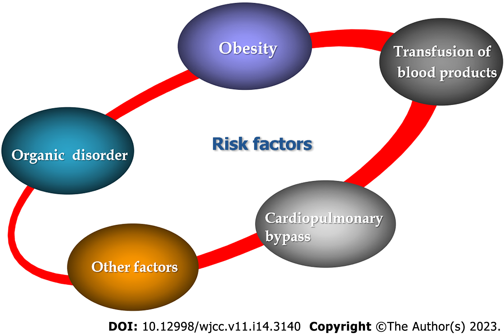
Hypoxemia is one of the major complications after surgery for Stanford type A aortic dissection. Comprehensively, the pathogenesis and development of postoperative hypoxemia involve the interaction of many risk factors including organic disorder, obesity, transfusion, and CPB (Figure 1). For treatment, the combination between medicine and supportive intervention is considered the more sustainable model. Surgically, all perioperative points should be managed with caution and patience, starting with reasonable preoperative lung function assessment, experienced cooperation of surgical team, and flawless postoperative rehabilitation. The vision, therefore, seeks to have larger scale studies concentrating on the long-term outcome of postoperative hypoxemia and more effective and optimal management.

Figure 1
Figure 1 Risk factors of postoperative hypoxemia for patients with Stanford type A aortic dissection.
Footnotes

Provenance and peer review: Unsolicited article; Externally peer reviewed.

Peer-review model: Single blind

Specialty type: Medicine, research and experimental

Country/Territory of origin: China

Peer-review report’s scientific quality classification

Grade A (Excellent): 0

Grade B (Very good): B

Grade C (Good): C

Grade D (Fair): 0

Grade E (Poor): 0

P-Reviewer: Papazafiropoulou A, Greece; Ueda H, Japan S-Editor: Fan JR L-Editor: Filipodia P-Editor: Li X

References
1.  Sayed A, Munir M, Bahbah EI. Aortic Dissection: A Review of the Pathophysiology, Management and Prospective Advances. Curr Cardiol Rev. 2021;17:e230421186875.  [PubMed]  [DOI]  [Cited in This Article: ]  [Cited by in Crossref: 5]  [Cited by in F6Publishing: 63]  [Article Influence: 15.8]  [Reference Citation Analysis (0)]
2.  Gu J, Chen Z. Clinical Efficacy of Hybrid Surgery for Stanford Type A Aortic Dissection. Risk Manag Healthc Policy. 2021;14:3013-3023.  [PubMed]  [DOI]  [Cited in This Article: ]  [Cited by in Crossref: 2]  [Reference Citation Analysis (0)]
3.  Jormalainen M, Kesävuori R, Raivio P, Vento A, Mustonen C, Honkanen HP, Rosato S, Simpanen J, Teittinen K, Biancari F, Juvonen T. Long-term outcomes after ascending aortic replacement and aortic root replacement for type A aortic dissection. Interact Cardiovasc Thorac Surg. 2022;34:453-461.  [PubMed]  [DOI]  [Cited in This Article: ]  [Cited by in Crossref: 1]  [Cited by in F6Publishing: 5]  [Article Influence: 1.3]  [Reference Citation Analysis (0)]
4.  Zhang L, Zhang L, Zhao Z, Liu Y, Wang J, Niu M, Sun X, Zhao X. Metabolic syndrome and its components are associated with hypoxemia after surgery for acute type A aortic dissection: an observational study. J Cardiothorac Surg. 2022;17:151.  [PubMed]  [DOI]  [Cited in This Article: ]  [Cited by in F6Publishing: 7]  [Reference Citation Analysis (0)]
5.  Sheng W, Le S, Song Y, Du Y, Wu J, Tang C, Wang H, Chen X, Wang S, Luo J, Li R, Xia J, Huang X, Ye P, Wu L, Du X, Wang D. Preoperative Nomogram and Risk Calculator for Postoperative Hypoxemia and Related Clinical Outcomes Following Stanford Type A Acute Aortic Dissection Surgery. Front Cardiovasc Med. 2022;9:851447.  [PubMed]  [DOI]  [Cited in This Article: ]  [Cited by in Crossref: 2]  [Cited by in F6Publishing: 2]  [Article Influence: 0.7]  [Reference Citation Analysis (0)]
6.  Guo Z, Yang Y, Zhao M, Zhang B, Lu J, Jin M, Cheng W. Preoperative hypoxemia in patients with type A acute aortic dissection: a retrospective study on incidence, related factors and clinical significance. J Thorac Dis. 2019;11:5390-5397.  [PubMed]  [DOI]  [Cited in This Article: ]  [Cited by in Crossref: 9]  [Cited by in F6Publishing: 16]  [Article Influence: 2.7]  [Reference Citation Analysis (0)]
7.  Wang Y, Xue S, Zhu H. Risk factors for postoperative hypoxemia in patients undergoing Stanford A aortic dissection surgery. J Cardiothorac Surg. 2013;8:118.  [PubMed]  [DOI]  [Cited in This Article: ]  [Cited by in Crossref: 21]  [Cited by in F6Publishing: 37]  [Article Influence: 3.1]  [Reference Citation Analysis (0)]
8.  Tsuruda T, Yamashita A, Otsu M, Koide M, Nakamichi Y, Sekita-Hatakeyama Y, Hatakeyama K, Funamoto T, Chosa E, Asada Y, Udagawa N, Kato J, Kitamura K. Angiotensin II Induces Aortic Rupture and Dissection in Osteoprotegerin-Deficient Mice. J Am Heart Assoc. 2022;11:e025336.  [PubMed]  [DOI]  [Cited in This Article: ]  [Cited by in F6Publishing: 6]  [Reference Citation Analysis (0)]
9.  Wu Z, Dai F, Ren W, Liu H, Li B, Chang J. Angiotensin II induces apoptosis of human pulmonary microvascular endothelial cells in acute aortic dissection complicated with lung injury patients through modulating the expression of monocyte chemoattractant protein-1. Am J Transl Res. 2016;8:28-36.  [PubMed]  [DOI]  [Cited in This Article: ]
10.  Gong M, Wu Z, Xu S, Li L, Wang X, Guan X, Zhang H. Increased risk for the development of postoperative severe hypoxemia in obese women with acute type a aortic dissection. J Cardiothorac Surg. 2019;14:81.  [PubMed]  [DOI]  [Cited in This Article: ]  [Cited by in Crossref: 26]  [Cited by in F6Publishing: 22]  [Article Influence: 3.7]  [Reference Citation Analysis (0)]
11.  Sheng W, Yang HQ, Chi YF, Niu ZZ, Lin MS, Long S. Independent risk factors for hypoxemia after surgery for acute aortic dissection. Saudi Med J. 2015;36:940-946.  [PubMed]  [DOI]  [Cited in This Article: ]  [Cited by in Crossref: 11]  [Cited by in F6Publishing: 23]  [Article Influence: 2.6]  [Reference Citation Analysis (0)]
12.  Shimizu T, Kimura N, Mieno M, Hori D, Shiraishi M, Tashima Y, Yuri K, Itagaki R, Aizawa K, Kawahito K, Yamaguchi A. Effects of Obesity on Outcomes of Acute Type A Aortic Dissection Repair in Japan. Circ Rep. 2020;2:639-647.  [PubMed]  [DOI]  [Cited in This Article: ]  [Cited by in Crossref: 1]  [Cited by in F6Publishing: 1]  [Article Influence: 0.2]  [Reference Citation Analysis (0)]
13.  Shi S, Gao Y, Wang L, Liu J, Yuan Z, Yu M. Elevated free fatty acid level is a risk factor for early postoperative hypoxemia after on-pump coronary artery bypass grafting: association with endothelial activation. J Cardiothorac Surg. 2015;10:122.  [PubMed]  [DOI]  [Cited in This Article: ]  [Cited by in Crossref: 9]  [Cited by in F6Publishing: 10]  [Article Influence: 1.0]  [Reference Citation Analysis (0)]
14.  Wu Z, Wang Z, Wu H, Hu R, Ren W, Hu Z, Chang J. Obesity is a risk factor for preoperative hypoxemia in Stanford A acute aortic dissection. Medicine (Baltimore). 2020;99:e19186.  [PubMed]  [DOI]  [Cited in This Article: ]  [Cited by in Crossref: 14]  [Cited by in F6Publishing: 24]  [Article Influence: 4.8]  [Reference Citation Analysis (0)]
15.  Wang D, Xu JZ, Kang YY, Zhang W, Hu LX, Wang JG. Aortic Root Diameter in Hypertensive Patients With Various Stages of Obstructive Sleep Apnea. Am J Hypertens. 2022;35:142-148.  [PubMed]  [DOI]  [Cited in This Article: ]  [Cited by in Crossref: 3]  [Cited by in F6Publishing: 3]  [Article Influence: 1.0]  [Reference Citation Analysis (0)]
16.  Maniaci A, Iannella G, Cocuzza S, Vicini C, Magliulo G, Ferlito S, Cammaroto G, Meccariello G, De Vito A, Nicolai A, Pace A, Artico M, Taurone S. Oxidative Stress and Inflammation Biomarker Expression in Obstructive Sleep Apnea Patients. J Clin Med. 2021;10.  [PubMed]  [DOI]  [Cited in This Article: ]  [Cited by in Crossref: 56]  [Cited by in F6Publishing: 109]  [Article Influence: 27.3]  [Reference Citation Analysis (0)]
17.  Xi X, Chen Y, Ma WG, Xie J, Liu YM, Zhu JM, Gong M, Zhu GF, Sun LZ. Is obstructive sleep apnoea associated with hypoxaemia and prolonged ICU stay after type A aortic dissection repair? BMC Cardiovasc Disord. 2021;21:421.  [PubMed]  [DOI]  [Cited in This Article: ]  [Reference Citation Analysis (0)]
18.  Klanderman RB, van Mourik N, Eggermont D, Peters AL, Tuinman PR, Bosman R, Endeman H, Cremer OL, Arbous SM, Vlaar APJ. Incidence of transfusion-related acute lung injury temporally associated with solvent/detergent plasma use in the ICU: A retrospective before and after implementation study. Transfusion. 2022;62:1752-1762.  [PubMed]  [DOI]  [Cited in This Article: ]  [Cited by in F6Publishing: 5]  [Reference Citation Analysis (0)]
19.  Yu Y, Jiang P, Sun P, Su N, Lin F. Pulmonary coagulation and fibrinolysis abnormalities that favor fibrin deposition in the lungs of mouse antibody-mediated transfusion-related acute lung injury. Mol Med Rep. 2021;24.  [PubMed]  [DOI]  [Cited in This Article: ]  [Cited by in Crossref: 2]  [Cited by in F6Publishing: 2]  [Article Influence: 0.5]  [Reference Citation Analysis (0)]
20.  Le A, Wu Y, Liu W, Wu C, Hu P, Zou J, Kuang L. MiR-144-induced KLF2 inhibition and NF-kappaB/CXCR1 activation promote neutrophil extracellular trap-induced transfusion-related acute lung injury. J Cell Mol Med. 2021;25:6511-6523.  [PubMed]  [DOI]  [Cited in This Article: ]  [Cited by in Crossref: 2]  [Cited by in F6Publishing: 13]  [Article Influence: 3.3]  [Reference Citation Analysis (0)]
21.  Bayat B, Nielsen KR, Bein G, Traum A, Burg-Roderfeld M, Sachs UJ. Transfusion of target antigens to preimmunized recipients: a new mechanism in transfusion-related acute lung injury. Blood Adv. 2021;5:3975-3985.  [PubMed]  [DOI]  [Cited in This Article: ]  [Cited by in Crossref: 5]  [Cited by in F6Publishing: 9]  [Article Influence: 2.3]  [Reference Citation Analysis (0)]
22.  Su IL, Wu VC, Chou AH, Yang CH, Chu PH, Liu KS, Tsai FC, Lin PJ, Chang CH, Chen SW. Risk factor analysis of postoperative acute respiratory distress syndrome after type A aortic dissection repair surgery. Medicine (Baltimore). 2019;98:e16303.  [PubMed]  [DOI]  [Cited in This Article: ]  [Cited by in Crossref: 13]  [Cited by in F6Publishing: 16]  [Article Influence: 2.7]  [Reference Citation Analysis (0)]
23.  He Y, Zhang HS, Zhang TZ, Feng Y, Zhu Y, Fan X. Analysis of the risk factors for severe lung injury after radical surgery for tetralogy of fallot. Front Surg. 2022;9:892562.  [PubMed]  [DOI]  [Cited in This Article: ]  [Reference Citation Analysis (0)]
24.  Deng Y, Hou L, Xu Q, Liu Q, Pan S, Gao Y, Dixon RAF, He Z, Wang X. Cardiopulmonary Bypass Induces Acute Lung Injury via the High-Mobility Group Box 1/Toll-Like Receptor 4 Pathway. Dis Markers. 2020;2020:8854700.  [PubMed]  [DOI]  [Cited in This Article: ]  [Cited by in Crossref: 4]  [Cited by in F6Publishing: 4]  [Article Influence: 0.8]  [Reference Citation Analysis (0)]
25.  Li J, Gao PF, Xu YX, Gu H, Wang QX. Probiotic Saccharomyces boulardii attenuates cardiopulmonary bypass-induced acute lung injury by inhibiting ferroptosis. Am J Transl Res. 2022;14:5003-5013.  [PubMed]  [DOI]  [Cited in This Article: ]
26.  Kong M, Wei D, Li X, Zhu X, Hong Z, Ni M, Wang Y, Dong A. The dynamic changes in autophagy activity and its role in lung injury after deep hypothermic circulatory arrest. J Cell Mol Med. 2022;26:1113-1127.  [PubMed]  [DOI]  [Cited in This Article: ]  [Cited by in Crossref: 1]  [Cited by in F6Publishing: 7]  [Article Influence: 2.3]  [Reference Citation Analysis (0)]
27.  Cooney SJ, Klawitter J, Khailova L, Robison J, Jaggers J, Ing RJ, Lawson S, Frank BS, Lujan SO, Davidson JA. Regional lung metabolic profile in a piglet model of cardiopulmonary bypass with circulatory arrest. Metabolomics. 2021;17:89.  [PubMed]  [DOI]  [Cited in This Article: ]  [Cited by in Crossref: 3]  [Cited by in F6Publishing: 1]  [Article Influence: 0.3]  [Reference Citation Analysis (0)]
28.  Andualem AA, Yesuf KA. Incidence and associated factors of postoperative hypoxemia among adult elective surgical patients at Dessie Comprehensive Specialized Hospital: An observational study. Ann Med Surg (Lond). 2022;78:103747.  [PubMed]  [DOI]  [Cited in This Article: ]  [Cited by in Crossref: 1]  [Reference Citation Analysis (0)]
29.  Kaushal A, Goyal P, Dhiraaj S, Agarwal A, Singh PK. Identification of Various Perioperative Risk Factors Responsible for Development of Postoperative Hypoxaemia. Turk J Anaesthesiol Reanim. 2018;46:416-423.  [PubMed]  [DOI]  [Cited in This Article: ]  [Cited by in Crossref: 7]  [Cited by in F6Publishing: 8]  [Article Influence: 1.1]  [Reference Citation Analysis (0)]
30.  Wu CP, Huang KL, Peng CK, Lan CC. Acute Hyperglycemia Aggravates Lung Injury via Activation of the SGK1-NKCC1 Pathway. Int J Mol Sci. 2020;21.  [PubMed]  [DOI]  [Cited in This Article: ]  [Cited by in Crossref: 8]  [Cited by in F6Publishing: 14]  [Article Influence: 2.8]  [Reference Citation Analysis (0)]
31.  Morey M, O'Gaora P, Pandit A, Hélary C. Hyperglycemia acts in synergy with hypoxia to maintain the pro-inflammatory phenotype of macrophages. PLoS One. 2019;14:e0220577.  [PubMed]  [DOI]  [Cited in This Article: ]  [Cited by in Crossref: 29]  [Cited by in F6Publishing: 55]  [Article Influence: 9.2]  [Reference Citation Analysis (0)]
32.  Jiang YX, Huang ZW. Ulinastatin alleviates pulmonary edema by reducing pulmonary permeability and stimulating alveolar fluid clearance in a rat model of acute lung injury. Iran J Basic Med Sci. 2022;25:1002-1008.  [PubMed]  [DOI]  [Cited in This Article: ]  [Cited by in F6Publishing: 2]  [Reference Citation Analysis (0)]
33.  Zhang G, Du Y, Sun N, Sun Y, Zhang L, Li X. Ulinastatin enhances autophagy against radiation-induced lung injury in mice. Transl Cancer Res. 2020;9:4162-4172.  [PubMed]  [DOI]  [Cited in This Article: ]  [Cited by in Crossref: 1]  [Cited by in F6Publishing: 1]  [Article Influence: 0.2]  [Reference Citation Analysis (0)]
34.  Zheng F, Wu X, Zhang J, Fu Z, Zhang Y. Sevoflurane reduces lipopolysaccharide-induced apoptosis and pulmonary fibrosis in the RAW264.7 cells and mice models to ameliorate acute lung injury by eliminating oxidative damages. Redox Rep. 2022;27:139-149.  [PubMed]  [DOI]  [Cited in This Article: ]  [Cited by in F6Publishing: 12]  [Reference Citation Analysis (0)]
35.  Fu Z, Wu X, Zheng F, Zhang Y. Sevoflurane anesthesia ameliorates LPS-induced acute lung injury (ALI) by modulating a novel LncRNA LINC00839/miR-223/NLRP3 axis. BMC Pulm Med. 2022;22:159.  [PubMed]  [DOI]  [Cited in This Article: ]  [Cited by in F6Publishing: 9]  [Reference Citation Analysis (0)]
36.  Zhang H, Liu Y, Meng X, Yang D, Shi S, Liu J, Yuan Z, Gu T, Han L, Lu F, Xu Z, Yu M. Effects of inhaled nitric oxide for postoperative hypoxemia in acute type A aortic dissection: a retrospective observational study. J Cardiothorac Surg. 2020;15:25.  [PubMed]  [DOI]  [Cited in This Article: ]  [Cited by in Crossref: 5]  [Cited by in F6Publishing: 7]  [Article Influence: 1.4]  [Reference Citation Analysis (0)]
37.  Zheng P, Jiang D, Liu C, Wei X, Li S. Nitric Oxide Inhalation Therapy Attenuates Postoperative Hypoxemia in Obese Patients with Acute Type A Aortic Dissection. Comput Math Methods Med. 2022;2022:9612548.  [PubMed]  [DOI]  [Cited in This Article: ]  [Cited by in Crossref: 4]  [Cited by in F6Publishing: 5]  [Article Influence: 1.7]  [Reference Citation Analysis (0)]
38.  Ma GG, Hao GW, Lai H, Yang XM, Liu L, Wang CS, Tu GW, Luo Z. Initial clinical impact of inhaled nitric oxide therapy for refractory hypoxemia following type A acute aortic dissection surgery. J Thorac Dis. 2019;11:495-504.  [PubMed]  [DOI]  [Cited in This Article: ]  [Cited by in Crossref: 7]  [Cited by in F6Publishing: 11]  [Article Influence: 1.8]  [Reference Citation Analysis (0)]
39.  Lin X, Ma X, Cui X, Zhang R, Pan H, Gao W. Effects of Erythropoietin on Lung Injury Induced by Cardiopulmonary Bypass After Cardiac Surgery. Med Sci Monit. 2020;26:e920039.  [PubMed]  [DOI]  [Cited in This Article: ]  [Cited by in Crossref: 3]  [Cited by in F6Publishing: 3]  [Article Influence: 0.6]  [Reference Citation Analysis (0)]
40.  Shi J, Yu T, Song K, Du S, He S, Hu X, Li X, Li H, Dong S, Zhang Y, Xie Z, Li C, Yu J. Dexmedetomidine ameliorates endotoxin-induced acute lung injury in vivo and in vitro by preserving mitochondrial dynamic equilibrium through the HIF-1a/HO-1 signaling pathway. Redox Biol. 2021;41:101954.  [PubMed]  [DOI]  [Cited in This Article: ]  [Cited by in Crossref: 69]  [Cited by in F6Publishing: 131]  [Article Influence: 32.8]  [Reference Citation Analysis (0)]
41.  Kim S, Park SJ, Nam SB, Song SW, Han Y, Ko S, Song Y. Pulmonary effects of dexmedetomidine infusion in thoracic aortic surgery under hypothermic circulatory arrest: a randomized placebo-controlled trial. Sci Rep. 2021;11:10975.  [PubMed]  [DOI]  [Cited in This Article: ]  [Cited by in Crossref: 6]  [Cited by in F6Publishing: 2]  [Article Influence: 0.5]  [Reference Citation Analysis (0)]
42.  Cao Z, Yang Z, Liang Z, Cen Q, Zhang Z, Liang H, Liu R, Zeng L, Xie Y, Wang Y. Prone vs Supine Position Ventilation in Adult Patients with Acute Respiratory Distress Syndrome: A Meta-Analysis of Randomized Controlled Trials. Emerg Med Int. 2020;2020:4973878.  [PubMed]  [DOI]  [Cited in This Article: ]  [Cited by in Crossref: 1]  [Cited by in F6Publishing: 1]  [Article Influence: 0.2]  [Reference Citation Analysis (0)]
43.  Aguirre-Bermeo H, Turella M, Bitondo M, Grandjean J, Italiano S, Festa O, Morán I, Mancebo J. Lung volumes and lung volume recruitment in ARDS: a comparison between supine and prone position. Ann Intensive Care. 2018;8:25.  [PubMed]  [DOI]  [Cited in This Article: ]  [Cited by in Crossref: 16]  [Cited by in F6Publishing: 17]  [Article Influence: 2.4]  [Reference Citation Analysis (0)]
44.  Xia WH, Yang CL, Chen Z, Ouyang CH, Ouyang GQ, Li QG. Clinical evaluation of prone position ventilation in the treatment of acute respiratory distress syndrome induced by sepsis. World J Clin Cases. 2022;10:5577-5585.  [PubMed]  [DOI]  [Cited in This Article: ]  [Cited by in CrossRef: 1]  [Cited by in F6Publishing: 1]  [Article Influence: 0.3]  [Reference Citation Analysis (0)]
45.  Fioretto JR, Klefens SO, Pires RF, Kurokawa CS, Carpi MF, Bonatto RC, Moraes MA, Ronchi CF. Comparison between conventional protective mechanical ventilation and high-frequency oscillatory ventilation associated with the prone position. Rev Bras Ter Intensiva. 2017;29:427-435.  [PubMed]  [DOI]  [Cited in This Article: ]  [Reference Citation Analysis (0)]
46.  Kiss T, Bluth T, Braune A, Huhle R, Denz A, Herzog M, Herold J, Vivona L, Millone M, Bergamaschi A, Andreeff M, Scharffenberg M, Wittenstein J, Vidal Melo MF, Koch T, Rocco PRM, Pelosi P, Kotzerke J, Gama de Abreu M. Effects of Positive End-Expiratory Pressure and Spontaneous Breathing Activity on Regional Lung Inflammation in Experimental Acute Respiratory Distress Syndrome. Crit Care Med. 2019;47:e358-e365.  [PubMed]  [DOI]  [Cited in This Article: ]  [Cited by in Crossref: 19]  [Cited by in F6Publishing: 24]  [Article Influence: 4.8]  [Reference Citation Analysis (0)]
47.  Wu X, Zheng R, Zhuang Z. Effect of transpulmonary pressure-guided positive end-expiratory pressure titration on lung injury in pigs with acute respiratory distress syndrome. J Clin Monit Comput. 2020;34:151-159.  [PubMed]  [DOI]  [Cited in This Article: ]  [Cited by in Crossref: 5]  [Cited by in F6Publishing: 5]  [Article Influence: 0.8]  [Reference Citation Analysis (0)]
48.  Boesing C, Graf PT, Schmitt F, Thiel M, Pelosi P, Rocco PRM, Luecke T, Krebs J. Effects of different positive end-expiratory pressure titration strategies during prone positioning in patients with acute respiratory distress syndrome: a prospective interventional study. Crit Care. 2022;26:82.  [PubMed]  [DOI]  [Cited in This Article: ]  [Cited by in Crossref: 2]  [Cited by in F6Publishing: 21]  [Article Influence: 7.0]  [Reference Citation Analysis (0)]
49.  Bime C, Fiero M, Lu Z, Oren E, Berry CE, Parthasarathy S, Garcia JGN. High Positive End-Expiratory Pressure Is Associated with Improved Survival in Obese Patients with Acute Respiratory Distress Syndrome. Am J Med. 2017;130:207-213.  [PubMed]  [DOI]  [Cited in This Article: ]  [Cited by in Crossref: 26]  [Cited by in F6Publishing: 27]  [Article Influence: 3.4]  [Reference Citation Analysis (0)]
50.  Pavlovsky B, Pesenti A, Spinelli E, Scaramuzzo G, Marongiu I, Tagliabue P, Spadaro S, Grasselli G, Mercat A, Mauri T. Effects of PEEP on regional ventilation-perfusion mismatch in the acute respiratory distress syndrome. Crit Care. 2022;26:211.  [PubMed]  [DOI]  [Cited in This Article: ]  [Cited by in F6Publishing: 7]  [Reference Citation Analysis (0)]
51.  Shang X, Wang Y. Comparison of outcomes of high-flow nasal cannula and noninvasive positive-pressure ventilation in patients with hypoxemia and various APACHE II scores after extubation. Ther Adv Respir Dis. 2021;15:17534666211004235.  [PubMed]  [DOI]  [Cited in This Article: ]  [Cited by in Crossref: 1]  [Cited by in F6Publishing: 7]  [Article Influence: 1.8]  [Reference Citation Analysis (0)]