©The Author(s) 2021.
World J Clin Cases. Feb 26, 2021; 9(6): 1461-1468
Published online Feb 26, 2021. doi: 10.12998/wjcc.v9.i6.1461
Published online Feb 26, 2021. doi: 10.12998/wjcc.v9.i6.1461
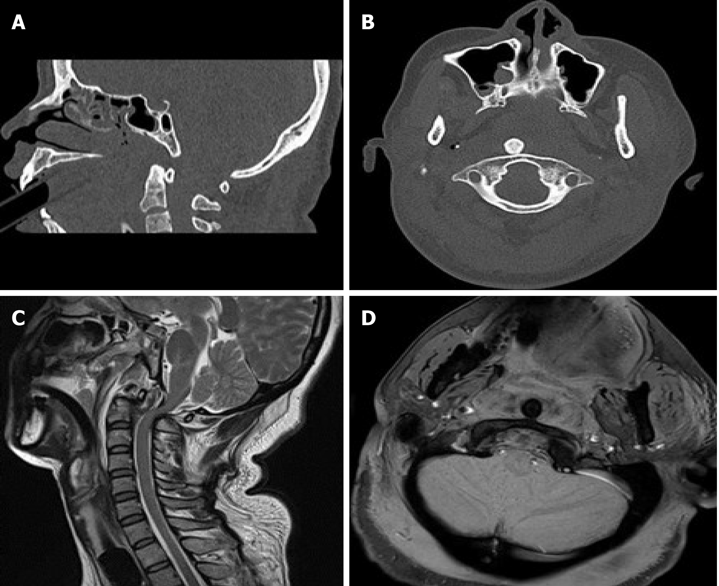
Figure 1 Radiographs on admission.
A and B: Computed tomography scans of midsagittal and coronal sections showing posterior atlantoaxial dislocation without fracture; C and D: T2W magnetic resonance imaging (MRI) midsagittal and T1W MRI coronal views showing signal changes with no cord edema but transverse ligament rupture.
Figure 2 Radiographs after closed reduction.
A: Lateral X-ray showing 11 mm atlantodental interval and 32 mm atlantoaxial interval of C1-C2; B and C: Computed tomography scans of coronal and midsagittal sections showing return of the odontoid process into the C1 ring with asymmetric lateral atlantodental spaces.
Figure 3 Radiographs after re-dislocation.
A: Lateral X-ray showing complete posterior dislocation of the atlantoaxial joint; B: Computed tomography scan of coronal section; C: Three-dimensional reconstruction showing C1-C2 re-dislocation without any associated fractures.
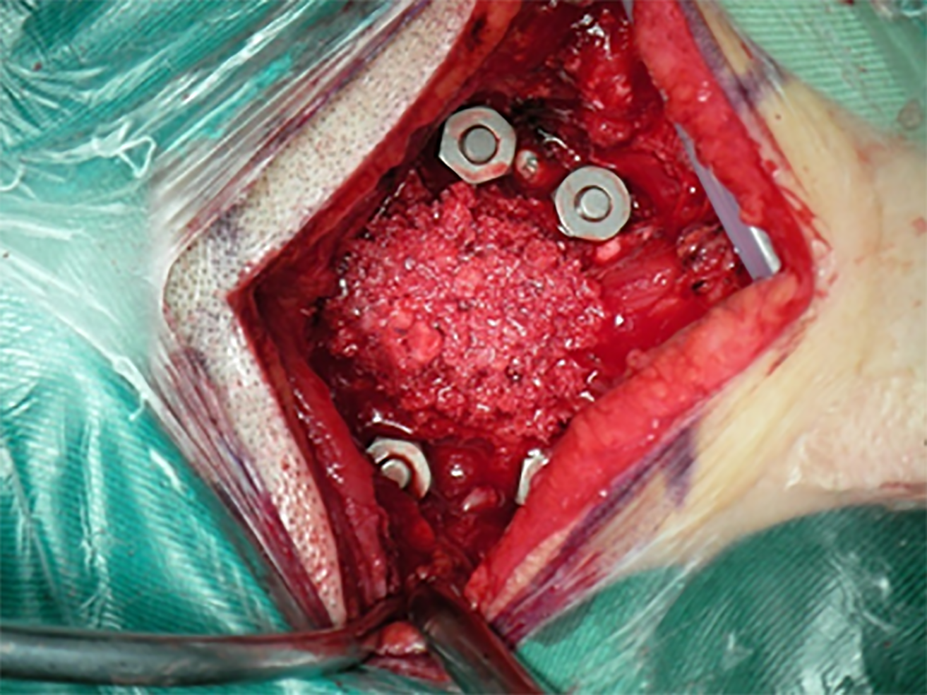
Figure 4 A cervical fixation system with four C1-C2 pedicular screws and autologous iliac bone grafts.
Figure 5 Radiographs 12 mo after internal fixation.
A: Lateral X-ray showing normal atlantoaxial complex, perfect positioning of pedicular screws, and good bone fusion; B: T2W magnetic resonance imaging (MRI) midsagittal; C: T1W MRI coronal views showing normal cervical spinal curvature and positioning of the odontoid process in the C1 ring without cord edema.
- Citation: Sun YH, Wang L, Ren JT, Wang SX, Jiao ZD, Fang J. Early reoccurrence of traumatic posterior atlantoaxial dislocation without fracture: A case report. World J Clin Cases 2021; 9(6): 1461-1468
- URL: https://www.wjgnet.com/2307-8960/full/v9/i6/1461.htm
- DOI: https://dx.doi.org/10.12998/wjcc.v9.i6.1461


