©The Author(s) 2021.
World J Clin Cases. Dec 16, 2021; 9(35): 11061-11070
Published online Dec 16, 2021. doi: 10.12998/wjcc.v9.i35.11061
Published online Dec 16, 2021. doi: 10.12998/wjcc.v9.i35.11061
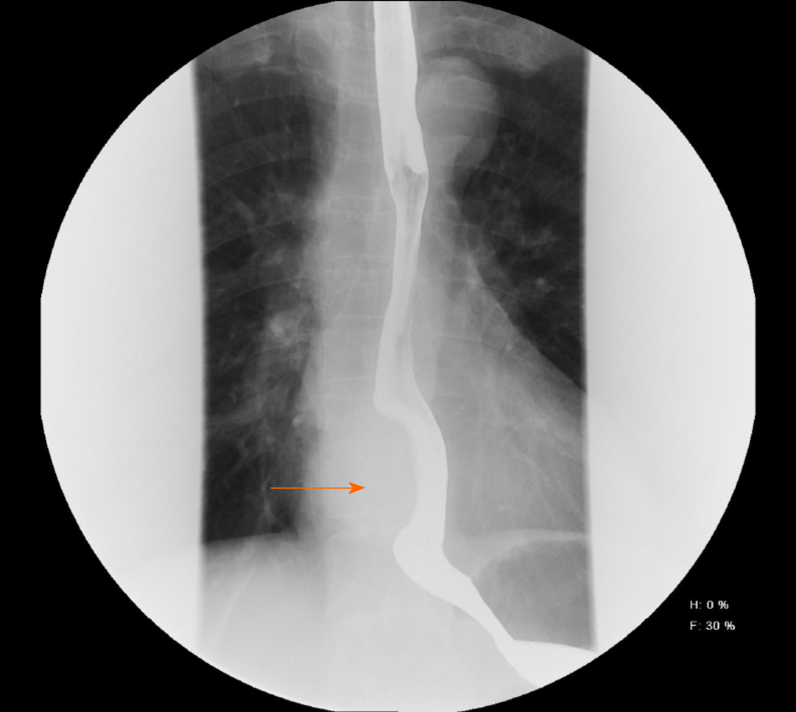
Figure 1 Barium esophagogram showed that the lower esophagus was compressed to approximately 5.
5 cm (arrowheads).
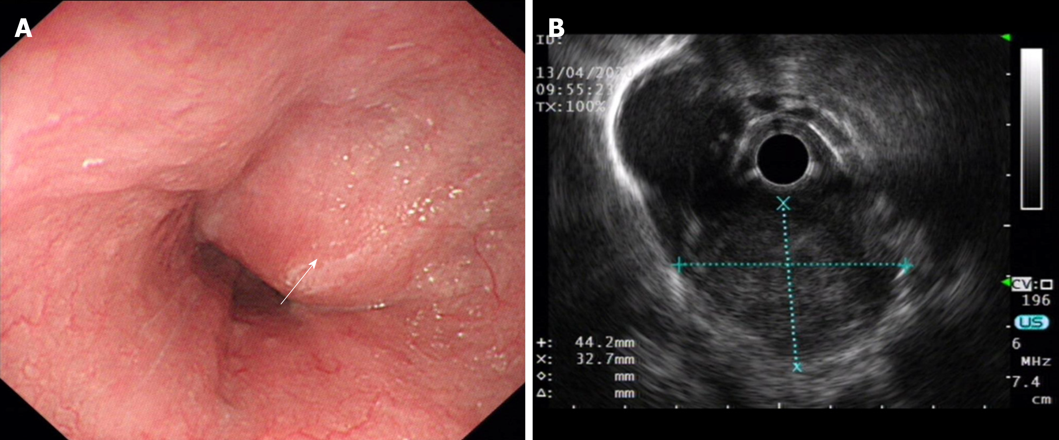
Figure 2 Endoscopy and endoscopic ultrasound image.
A: Esophagogastroduodenoscopy showed a large lesion in the esophagus 32-38 cm from the incisors (arrowheads); B: Endoscopic ultrasound showed a hypoechoic lesion, likely originating in the muscularis propria.
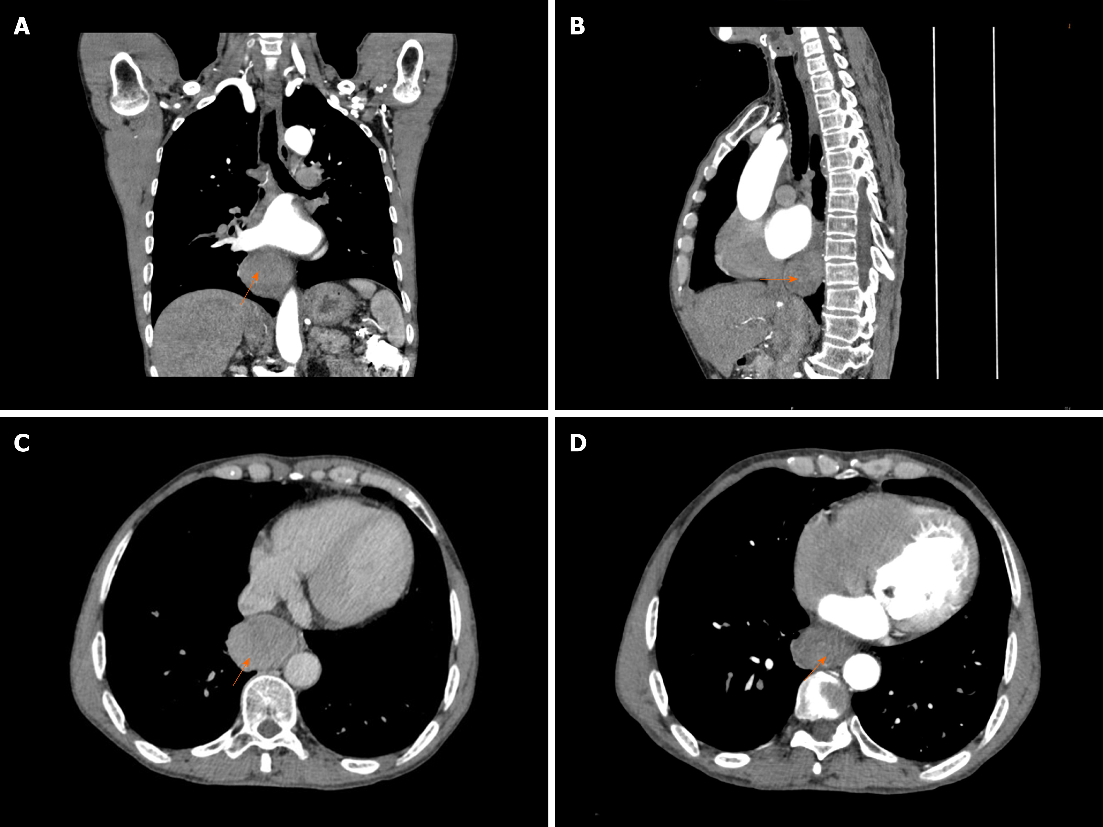
Figure 3 Chest computed tomography revealed a mass approximately 53 mm × 39 mm × 50 mm protruding from the lower esophagus (arrowheads).
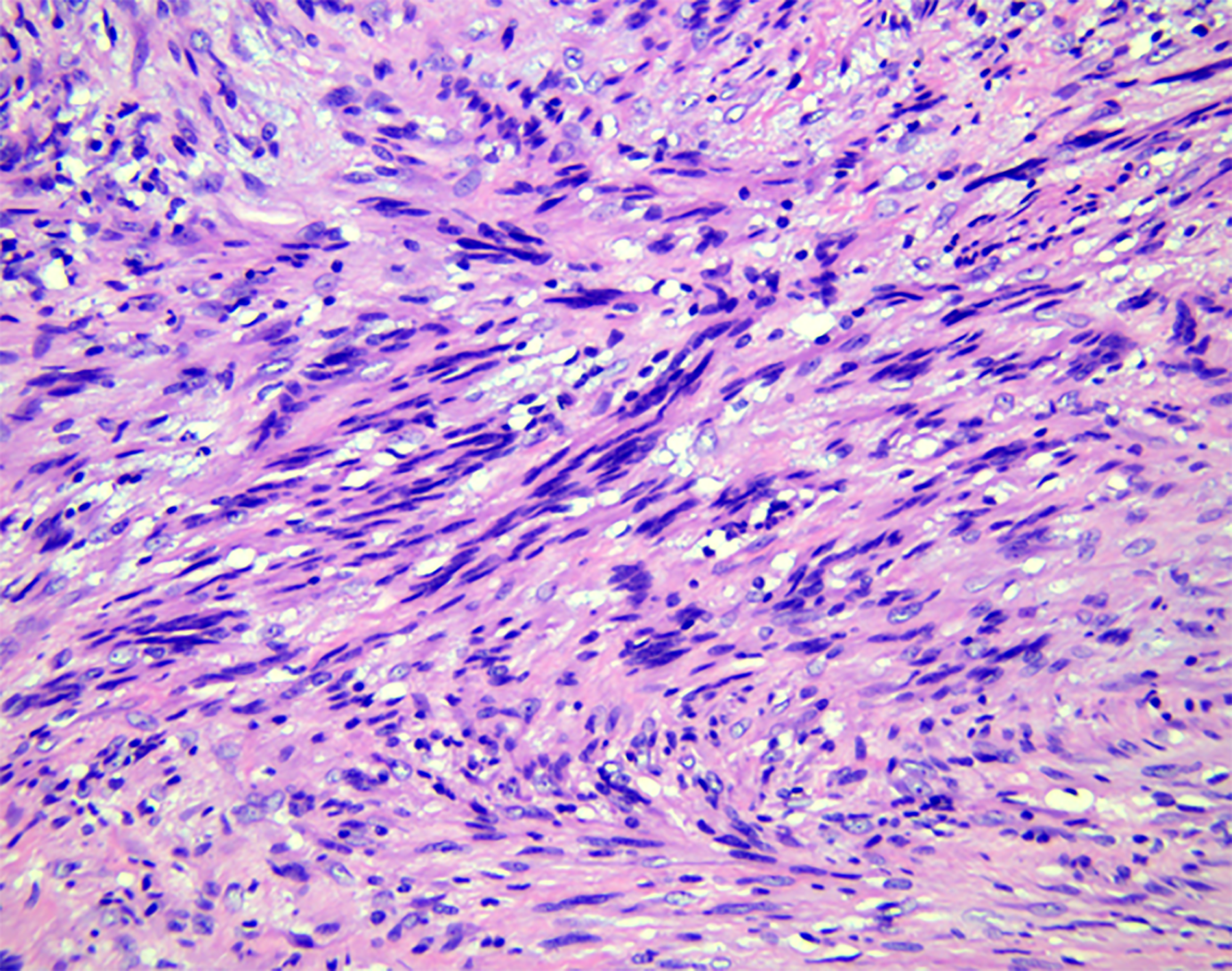
Figure 4 Histopathological examination revealed spindle-shaped cells arranged in interlacing fascicles (hematoxylin and eosin stain, × 200).
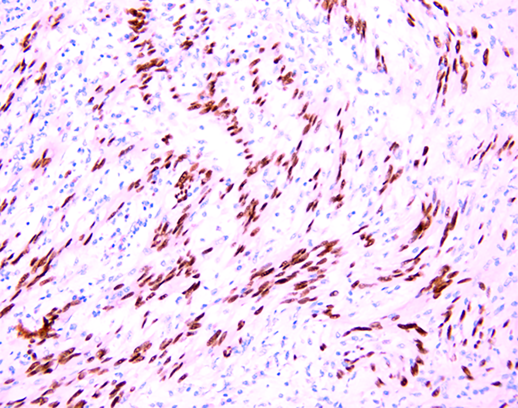
Figure 5 Immunohistochemical examination revealed SOX10 protein positivity (× 200).
- Citation: Wang TY, Wang BL, Wang FR, Jing MY, Zhang LD, Zhang DK. Thoracoscopic resection of a large lower esophageal schwannoma: A case report and review of the literature. World J Clin Cases 2021; 9(35): 11061-11070
- URL: https://www.wjgnet.com/2307-8960/full/v9/i35/11061.htm
- DOI: https://dx.doi.org/10.12998/wjcc.v9.i35.11061


