©The Author(s) 2021.
World J Clin Cases. Nov 16, 2021; 9(32): 9942-9947
Published online Nov 16, 2021. doi: 10.12998/wjcc.v9.i32.9942
Published online Nov 16, 2021. doi: 10.12998/wjcc.v9.i32.9942
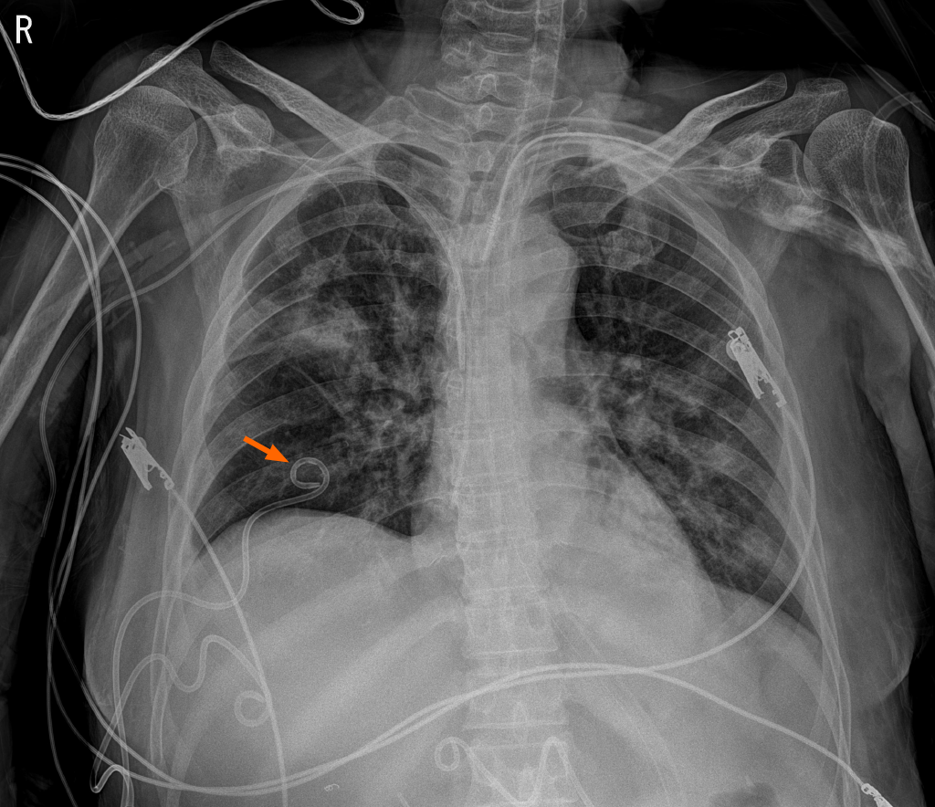
Figure 1
Portable chest radiography shows percutaneous catheter drainage for pleural effusion drain.
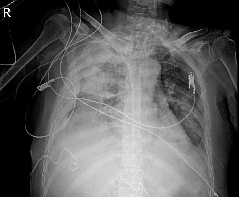
Figure 2
Portable chest radiography approximately 20 min after percutaneous catheter removal shows increased opacification of right hemithorax, and fluid is seen tracking up the lateral margin of the thorax.
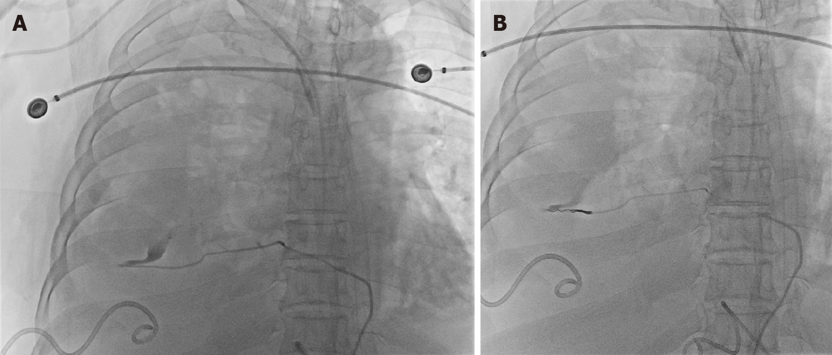
Figure 3 Transarterial thoracic aortography.
A: Extravasation of contrast is identified in the 7th intercostal artery distal portion on the right intercostal angiograms; B: After super-selection using a microcatheter, embolization is performed using gelfoam and microcoil.
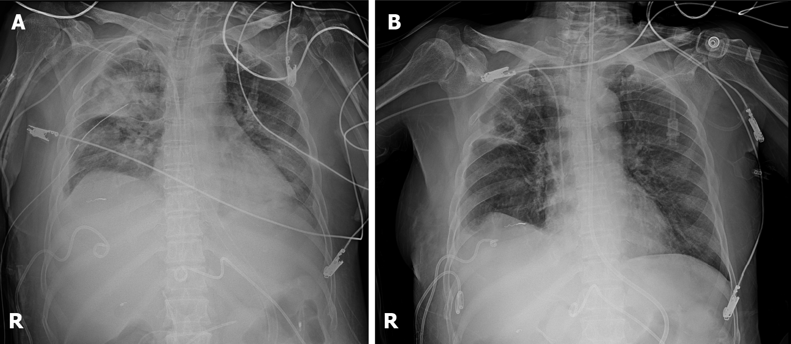
Figure 4 Portable chest radiography.
A: After 28-F chest tube is placed inside of the right pleural space for hemothorax drainage; B: 3 wk after right 7th intercostal artery embolization.
- Citation: Park C, Lee J. Massive hemothorax due to intercostal arterial bleeding after percutaneous catheter removal in a multiple-trauma patient: A case report. World J Clin Cases 2021; 9(32): 9942-9947
- URL: https://www.wjgnet.com/2307-8960/full/v9/i32/9942.htm
- DOI: https://dx.doi.org/10.12998/wjcc.v9.i32.9942


