©The Author(s) 2021.
World J Clin Cases. Nov 16, 2021; 9(32): 9935-9941
Published online Nov 16, 2021. doi: 10.12998/wjcc.v9.i32.9935
Published online Nov 16, 2021. doi: 10.12998/wjcc.v9.i32.9935
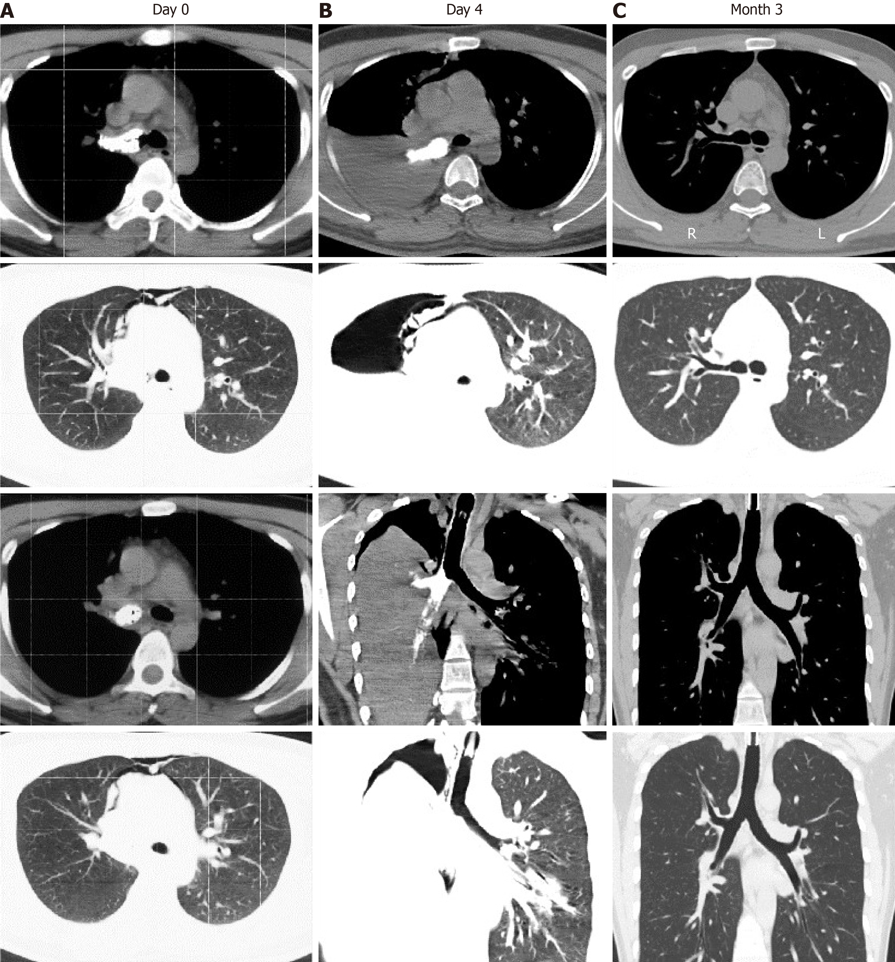
Figure 1 Computed tomography (CT) manifestation of the patient’s lung with lime aspiration.
A: CT after lime aspiration showed a high-intensity mass in the right principle bronchus, and pneumomediastinum; B: Follow-up CT at 1 d before bronchoscopy showed secondary right pulmonary atelectasis, and aeropleura; C: Follow-up CT at 3 mo showed anatomical recovery of the bronchus and lung.
Figure 2 Bronchoscopy of the bronchial bifurcation before and after lime clearance.
A: Before lime clearance, bronchoscopy showed the entry of the right principle bronchus was completely obstructed by lime. Hyperemia, edema and erosion were seen in the tracheal mucosa and left bronchus; B: After mechanical clearance of the lime, the right primary bronchus was reopened; C: Three months later, the mucosa at all bronchial levels was smooth and no stenosis or occlusion was found in the bronchus.
- Citation: Li XY, Hou HJ, Dai B, Tan W, Zhao HW. Adult with mass burnt lime aspiration: A case report and literature review. World J Clin Cases 2021; 9(32): 9935-9941
- URL: https://www.wjgnet.com/2307-8960/full/v9/i32/9935.htm
- DOI: https://dx.doi.org/10.12998/wjcc.v9.i32.9935


