©The Author(s) 2021.
World J Clin Cases. Nov 6, 2021; 9(31): 9635-9644
Published online Nov 6, 2021. doi: 10.12998/wjcc.v9.i31.9635
Published online Nov 6, 2021. doi: 10.12998/wjcc.v9.i31.9635
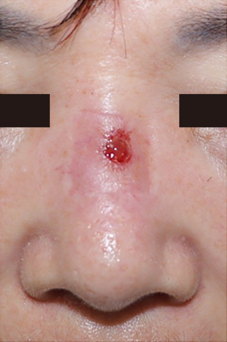
Figure 1 Photographic finding of case one’s initial visit.
Erythema and pus drainage with skin necrosis at the nasal dorsum were observed.
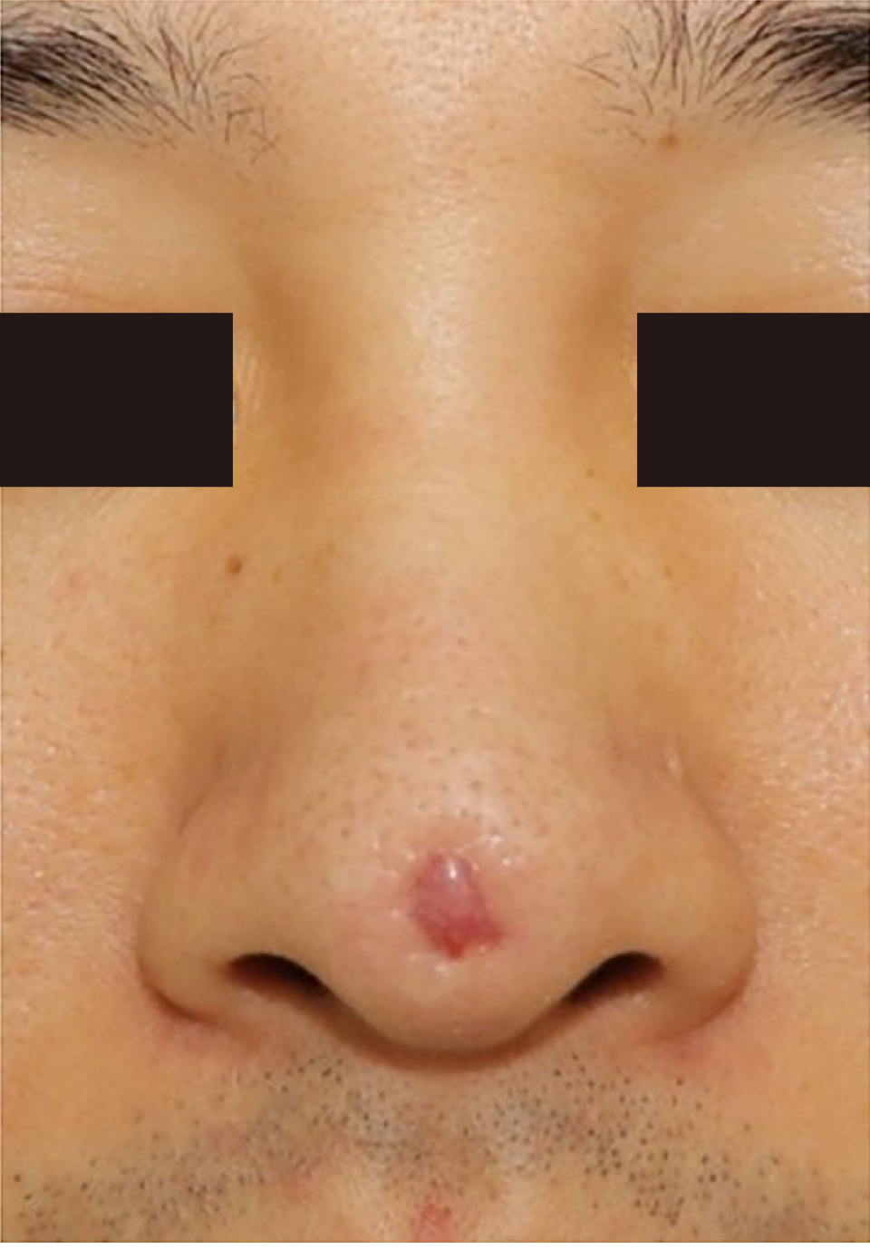
Figure 2 Photographic finding of patient’s initial visit.
Erythema with skin necrosis was observed at the nasal tip.
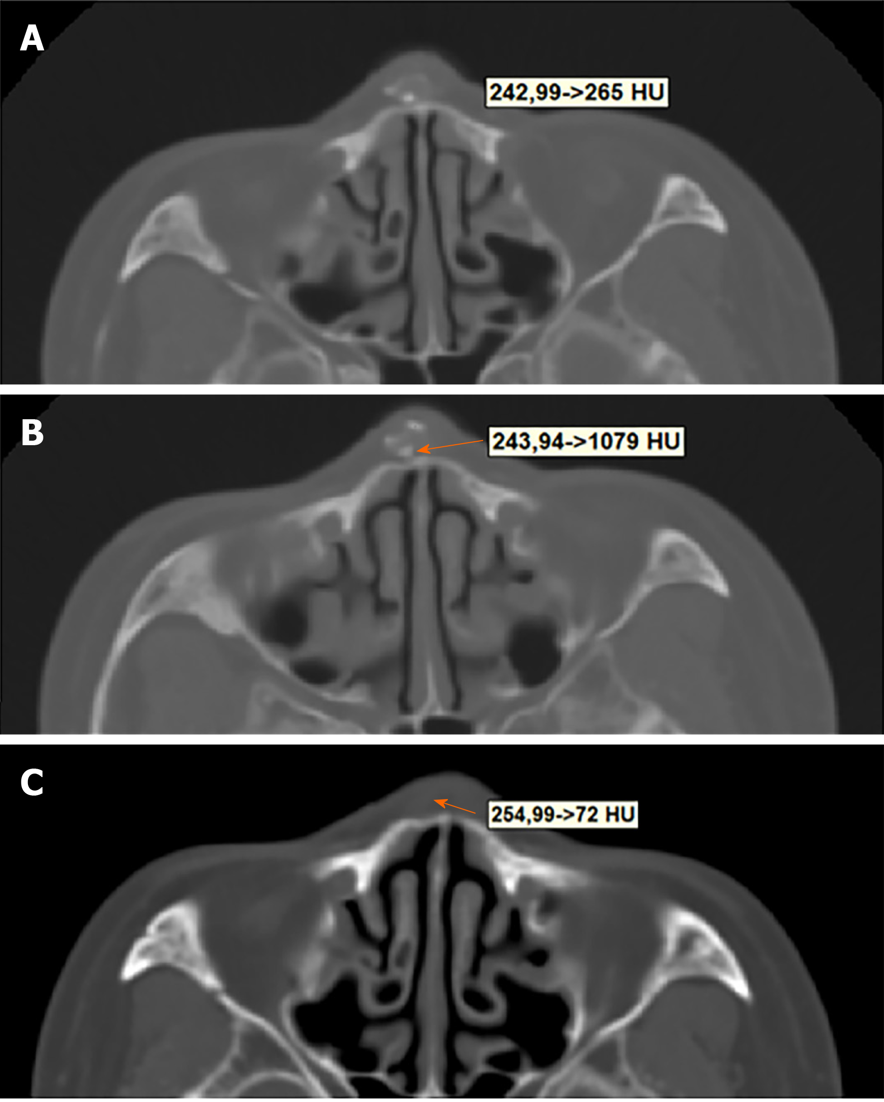
Figure 3 Hounsfield unit measurement of case one.
A: Inflammatory tissue; B: Calcified lesion estimated as polydioxanone (PDO); C: Inflammatory tissue at POD #5. It can be seen that the HU values decreased compared to the preoperative period. HU: Hounsfield unit.
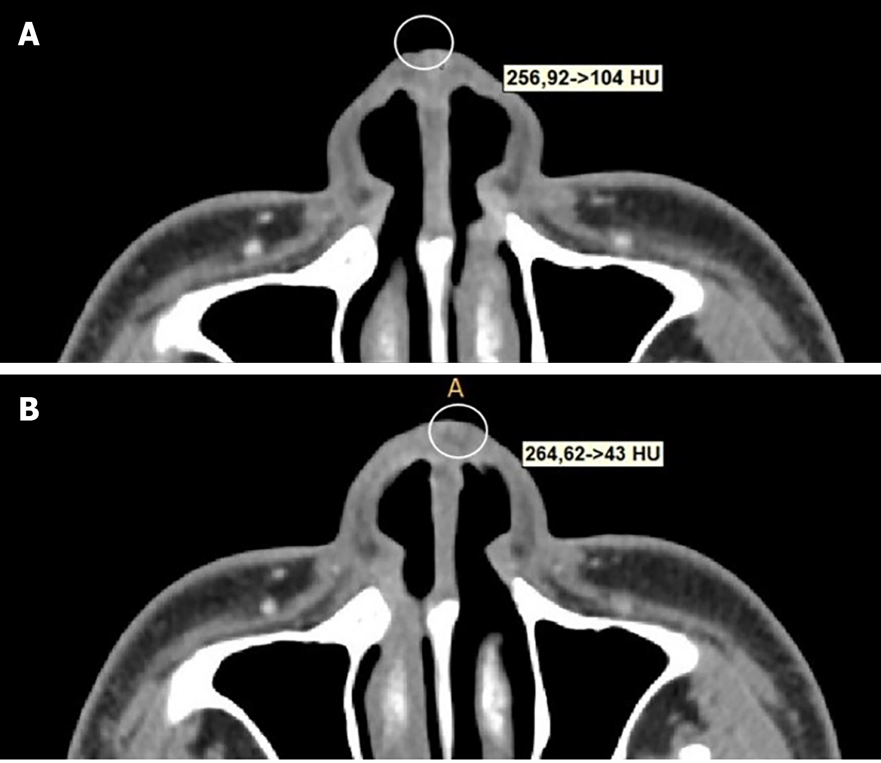
Figure 4 Hounsfield unit measurement of case two.
A: Preoperative measurement on suspected inflammatory tissue; B: Inflammatory tissue at polydioxanone #8.
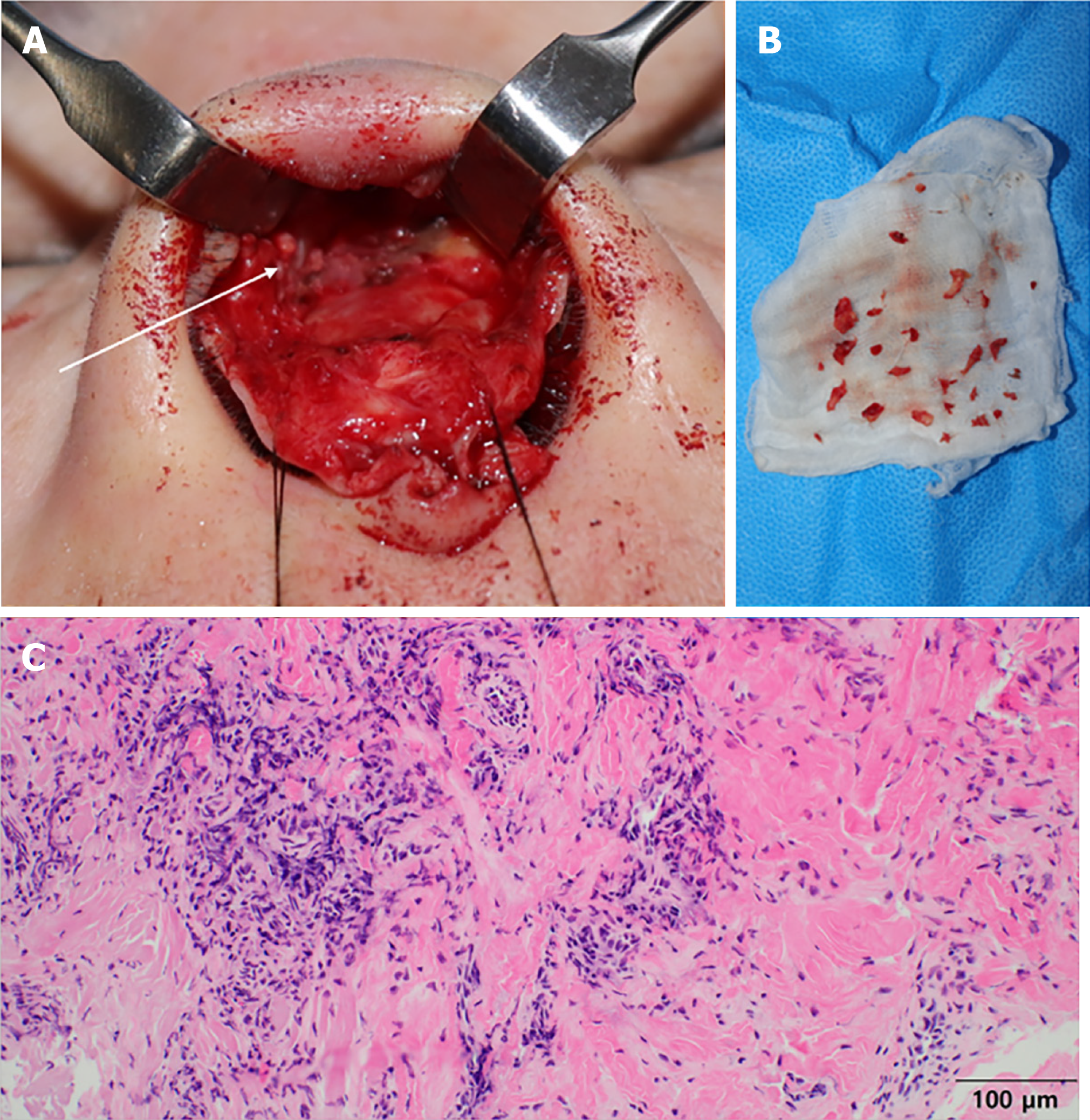
Figure 5 Clinical features of the patient one.
A: Intraoperative finding. capsule and foreign body granuloma (white arrow) around the polydioxanone (PDO) were observed in the sub-superficial musculoaponeurotic system layer; B: PDO fragment broken down into multiple pieces; C: Histopathological findings (Hematoxylin-eosin staining, magnification × 200).
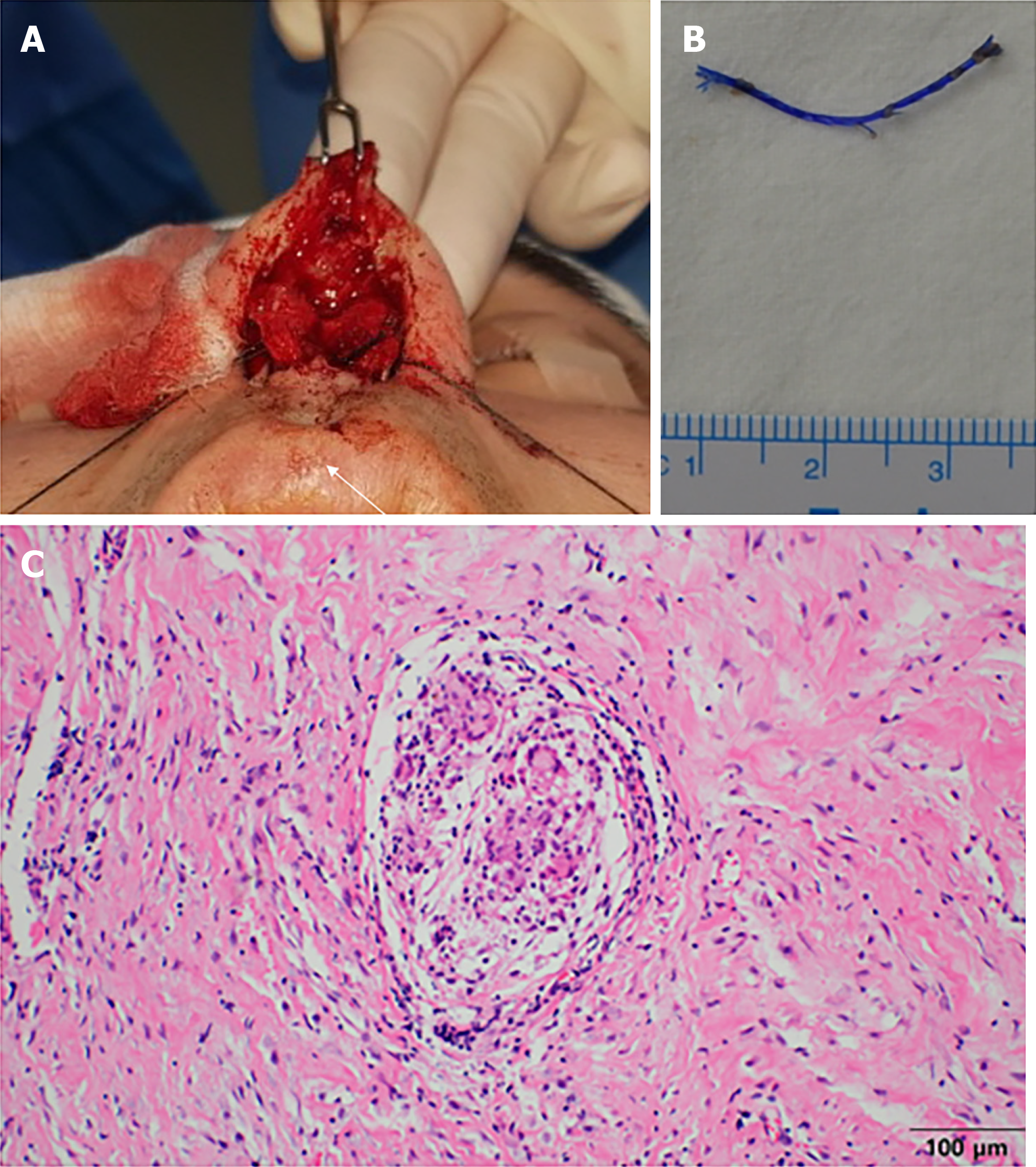
Figure 6 Clinical features of patient two.
A: Polypropylene threads with a barbed pattern located between medial crura of the lower lateral cartilage (white arrow); B: Removed barbed pattern thread; C: Histopathological findings (Hematoxylin-eosin staining, magnification × 200).
- Citation: Lee DW, Ryu H, Jang SH, Kim JH. Clinical features and literature review related to the material differences in thread rhinoplasty: Two case reports. World J Clin Cases 2021; 9(31): 9635-9644
- URL: https://www.wjgnet.com/2307-8960/full/v9/i31/9635.htm
- DOI: https://dx.doi.org/10.12998/wjcc.v9.i31.9635


