©The Author(s) 2021.
World J Clin Cases. Nov 6, 2021; 9(31): 9549-9556
Published online Nov 6, 2021. doi: 10.12998/wjcc.v9.i31.9549
Published online Nov 6, 2021. doi: 10.12998/wjcc.v9.i31.9549
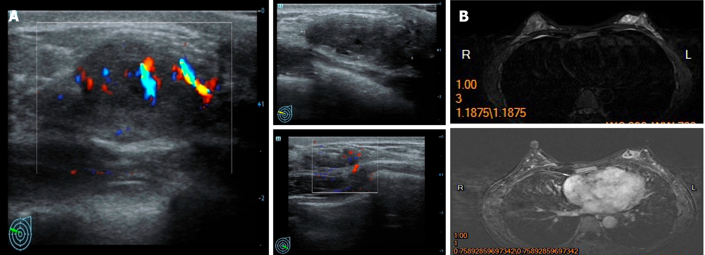
Figure 1 Imaging examinations in Case 1.
A: Mammary ultrasound revealed a 2.6 cm × 0.9 cm low echoic mass in her left breast; B: Left breast magnetic resonance imaging showed no invasion of the adjacent skin and nipple.
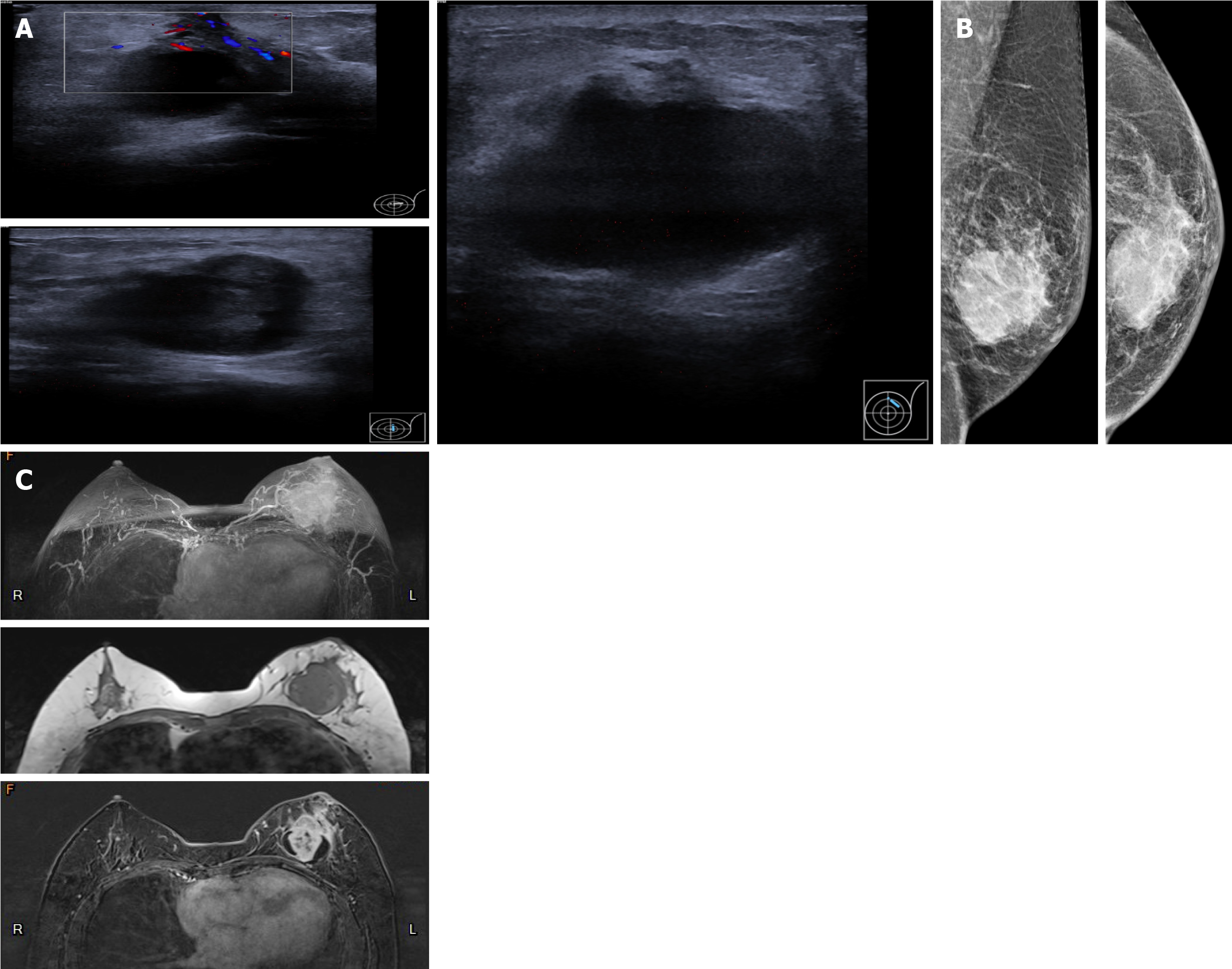
Figure 2 Imaging examinations in Case 2.
A: Ultrasound revealed a mixed echogenic mass of the left breast with ambiguous borders, measuring 4.0 cm × 2.6 cm × 4.5 cm; B: Mammography showed thickened peripheral skin and suspensory ligament; C: Magnetic resonance imaging showed a cystic lesion with irregular necrosis in the tumor.
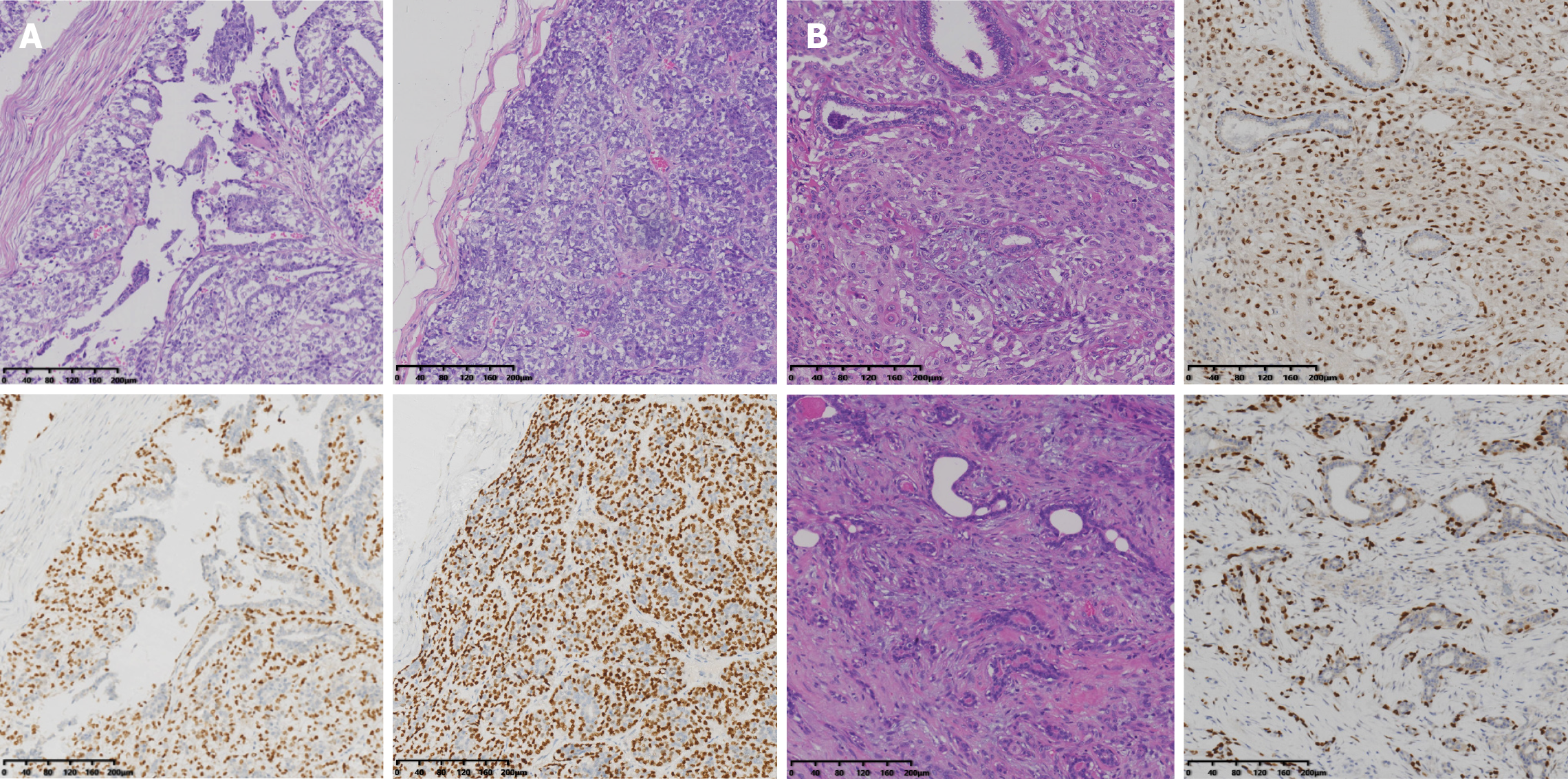
Figure 3 Pathological examinations.
A: Case 1; B: Case 2. Hematoxylin-eosin staining and immunohistochemical analysis for P63 staining (200 ×).
- Citation: Zhai DY, Zhen TT, Zhang XL, Luo J, Shi HJ, Shi YW, Shao N. Malignant adenomyoepithelioma of the breast: Two case reports and review of the literature. World J Clin Cases 2021; 9(31): 9549-9556
- URL: https://www.wjgnet.com/2307-8960/full/v9/i31/9549.htm
- DOI: https://dx.doi.org/10.12998/wjcc.v9.i31.9549


