©The Author(s) 2021.
World J Clin Cases. Nov 6, 2021; 9(31): 9535-9541
Published online Nov 6, 2021. doi: 10.12998/wjcc.v9.i31.9535
Published online Nov 6, 2021. doi: 10.12998/wjcc.v9.i31.9535
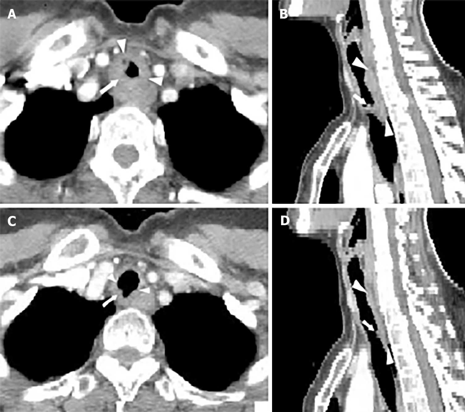
Figure 1 Imaging of the case.
A and B: Chest computed tomography pre-treatment revealed a soft tissue mass surrounding the inner surface of the upper trachea; C and D: Chest computed tomography after 60 Gy indicated dramatic regression of the tumor.
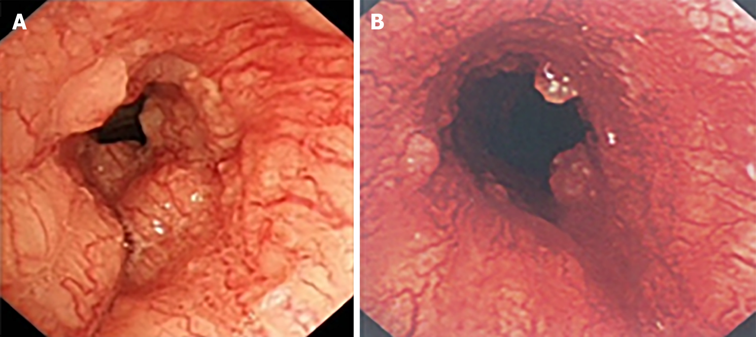
Figure 2 Bronchoscopic findings.
A: Pre-treatment revealed that a large polypoid intra-luminal mass, with its surface presenting rich vascular networks and nodular protrusions; B: After receiving 40 Gy, bronchoscopy indicated that the previous large polypoid intra-luminal mass was significantly eliminated.
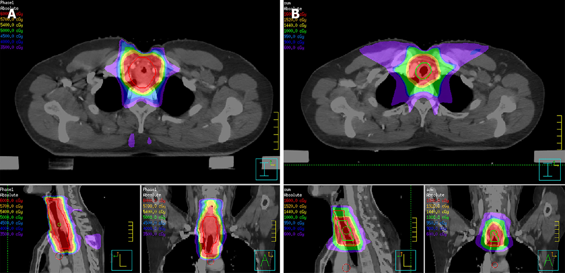
Figure 3 Dose distribution of the first and second treatment plan using intensity-modulated radiotherapy.
A: The first radiotherapy plan (30 × 2 Gy) with an irradiated high dose zone for CTVlow, surrounded by a red line; B: The second radiotherapy plan, where CTVhigh (red line) received an additional 10 Gy in 5 fractions and the GTV (green line) received another 6 Gy in 3 fractions. IMRT: Intensity modulated radiation therapy; CTV: Clinical target volume; GTV: Gross tumor volume.
- Citation: Wu Q, Xu F. Rapid response to radiotherapy in unresectable tracheal adenoid cystic carcinoma: A case report. World J Clin Cases 2021; 9(31): 9535-9541
- URL: https://www.wjgnet.com/2307-8960/full/v9/i31/9535.htm
- DOI: https://dx.doi.org/10.12998/wjcc.v9.i31.9535


