©The Author(s) 2021.
World J Clin Cases. Oct 26, 2021; 9(30): 9255-9268
Published online Oct 26, 2021. doi: 10.12998/wjcc.v9.i30.9255
Published online Oct 26, 2021. doi: 10.12998/wjcc.v9.i30.9255
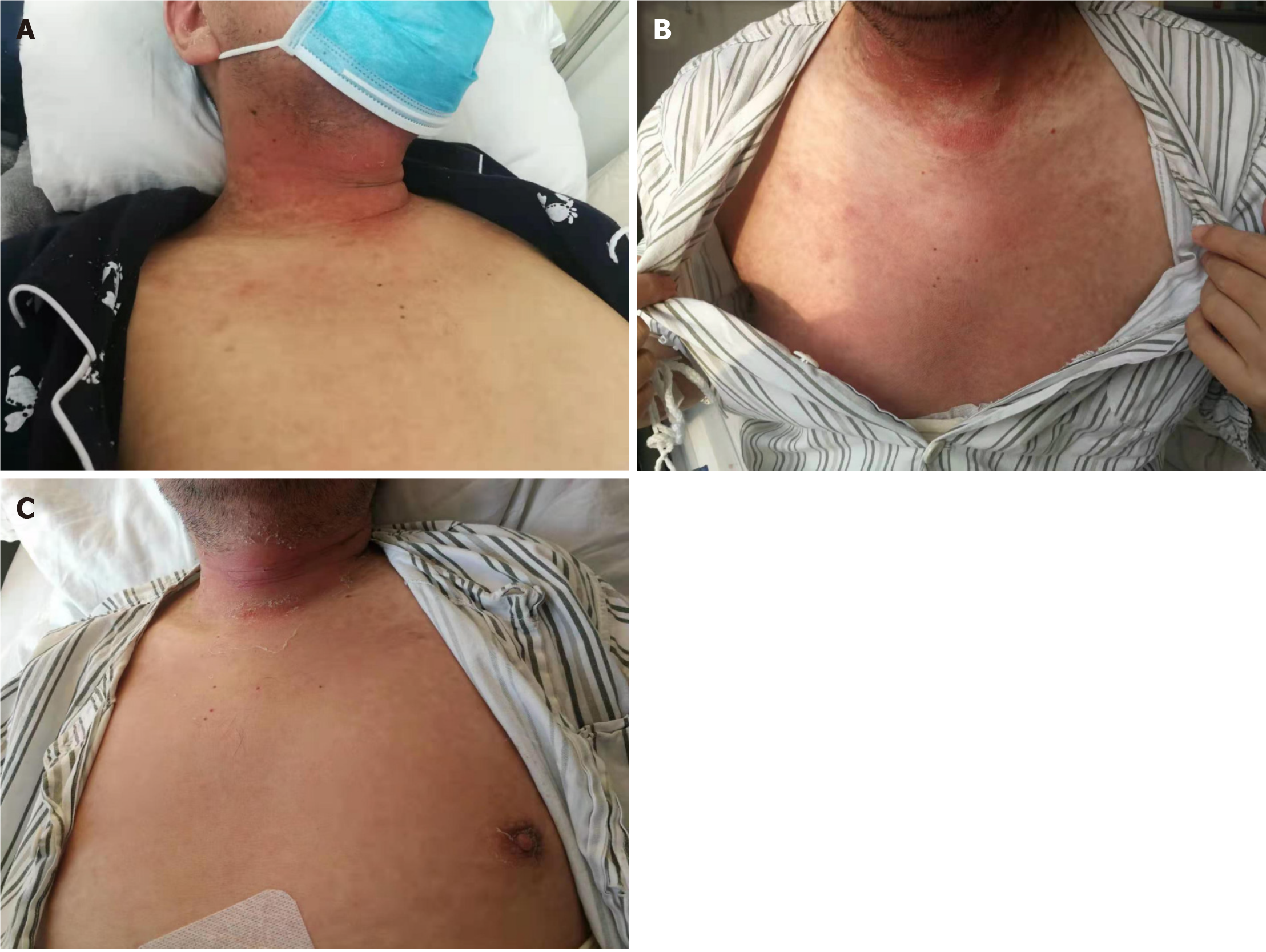
Figure 1 The skin rash evolution of acute graft-versus-host disease occurred in case 5.
A: On day 34 after liver transplantation (LT), he developed a maculo-papular skin rash on his palm, neck and upper extremities; B: On day 36 after LT, the rash progresses to the trunk, where it is partially fused into flakes, with extensive desquamation and flushing of the skin; C: On day 40 after LT, the skin rash was reduced and lighter in color, accompanied by extensive desquamation.
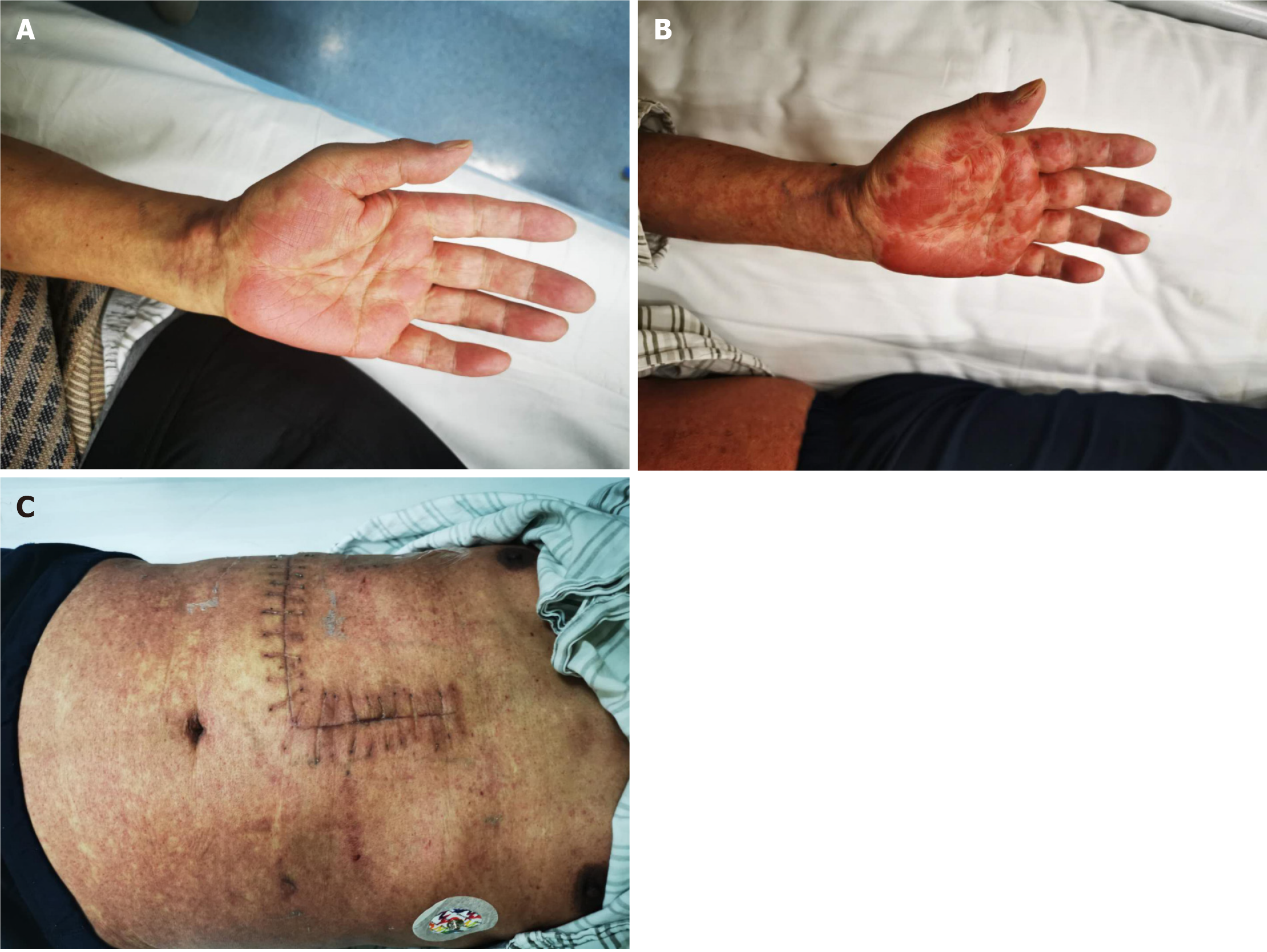
Figure 2 The skin rash evolution of acute graft-versus-host disease occurred in case 6.
A: On day 30 after liver transplantation (LT), he developed a maculo-papular skin rash on his palm, neck and limbs; B, C: On day 35 after LT, the skin rash gradually evolved into an erythematous maculopapular appearance, accompanied by partially confluent outbreak, it spread to the palm, face, neck, chest, and limbs.
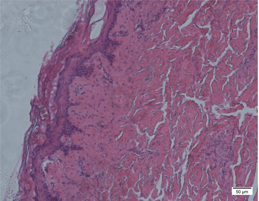
Figure 3 The abdominal skin biopsy showed that skin involvement with acute graft-versus-host disease in case 5 (Haematoxylin-eosin staining, HE × 200 times).
Small abdominal skin tissue, hyperkeratosis and dyskeratosis of the epidermis, lymphocyte infiltration around small vessels in the dermal line and around skin appendages, hyperplasia and sclerosis of the dermal fibrous tissue.
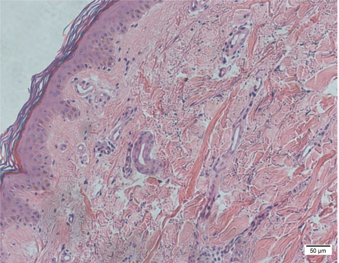
Figure 4 The right forearm skin biopsy showed that skin involvement with acute graft-versus-host disease in case 6 (Haematoxylin-eosin staining, HE × 200 times).
The right forearm skin tissue, the base layer of squamous epithelium is liquefied and degeneration. The localized area of the spinous cell layer was edema, focal necrosis and a few dyskeratosis cells were observed, and a few lymphocytes and other inflammatory cells were infiltrated in the superficial dermis.
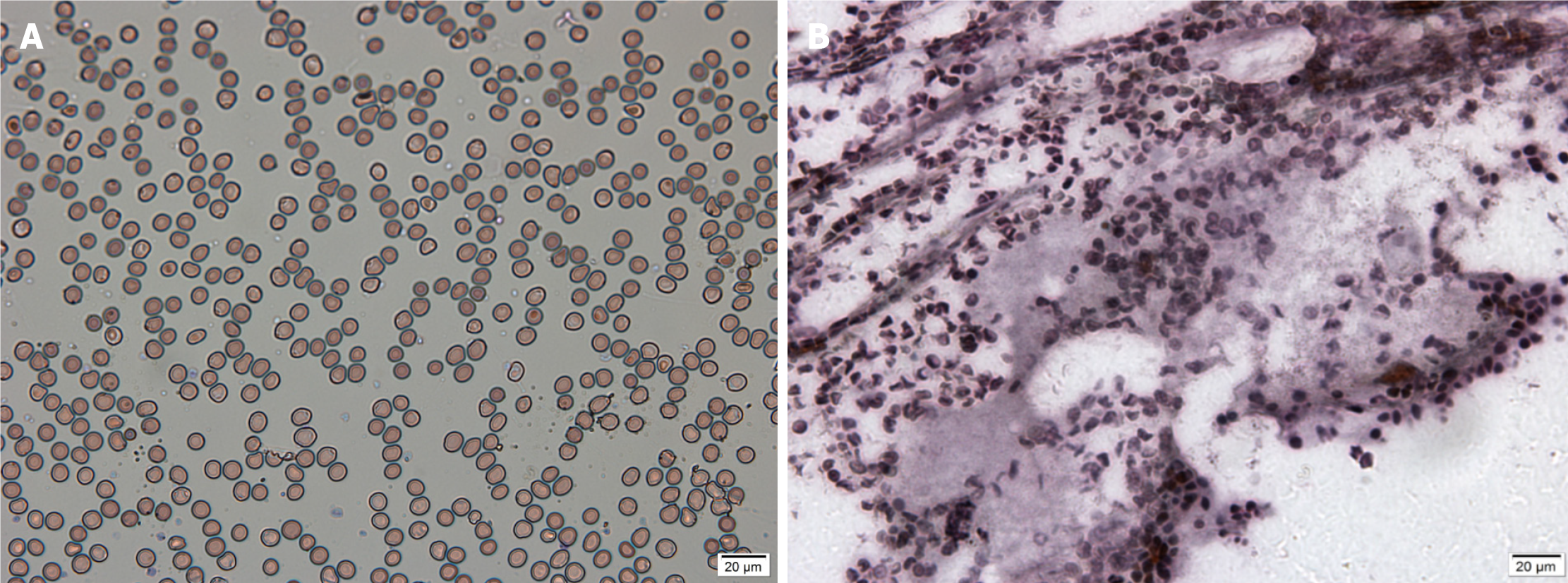
Figure 5 The bone marrow smear and bone marrow biopsy in case 3.
The images showed that the hyperplasia of bone marrow was extremely inhibited, the hyperplasia of granulocytes and erythrocytes was inhibited, lymphocytes accounted for 86%, and the morphology was generally normal. A (M × 400 times): Bone marrow smear; B (GO × 400 times): Bone marrow biopsy.
- Citation: Tian M, Lyu Y, Wang B, Liu C, Yu L, Shi JH, Liu XM, Zhang XG, Guo K, Li Y, Hu LS. Diagnosis and treatment of acute graft-versus-host disease after liver transplantation: Report of six cases. World J Clin Cases 2021; 9(30): 9255-9268
- URL: https://www.wjgnet.com/2307-8960/full/v9/i30/9255.htm
- DOI: https://dx.doi.org/10.12998/wjcc.v9.i30.9255


