©The Author(s) 2021.
World J Clin Cases. Oct 26, 2021; 9(30): 9182-9191
Published online Oct 26, 2021. doi: 10.12998/wjcc.v9.i30.9182
Published online Oct 26, 2021. doi: 10.12998/wjcc.v9.i30.9182
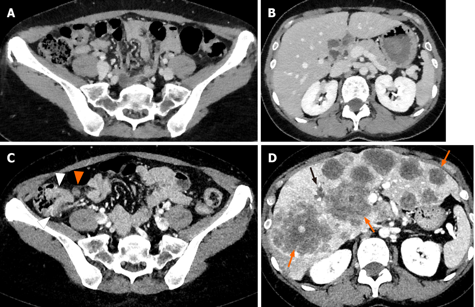
Figure 1 Computed tomography before corticosteroid administration and four months after the therapy.
A and B: No significant findings were noted before corticosteroid administration; C: Tumorous lesion in the ascending colon (white arrowheads) and suspicion of lymph node metastasis (orange arrowheads) were noted; D: A significant number of low-density masses in the liver up to 80 mm in size (orange arrows) were confirmed. Mild dilatation of the intrahepatic bile duct was noted (black arrow).
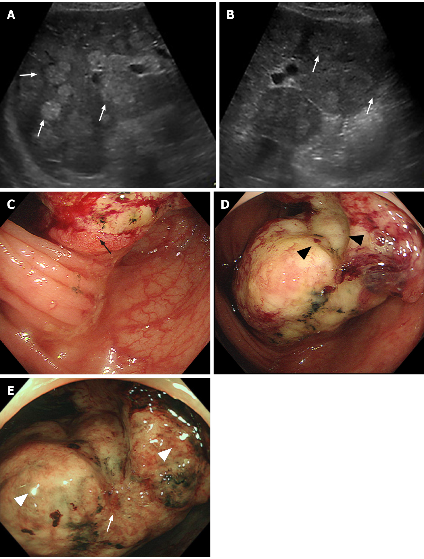
Figure 2 Ultrasonographic and endoscopic images of the tumor.
A and B: Abdominal ultrasonography revealed multiple iso- to high-echoic masses in bilateral liver lobes (white arrows). C-E: Endoscopic findings of the colon tumor. A large, solid, multinodular tumor on the epithelial layer (black arrow) with a semicircular depressive lesion at its center in the surface (black arrowheads) was observed in the ascending colon. The tumor was covered with whitish mucus and debris (white arrowheads), with an abnormal vascular structure on its surface (white arrows).
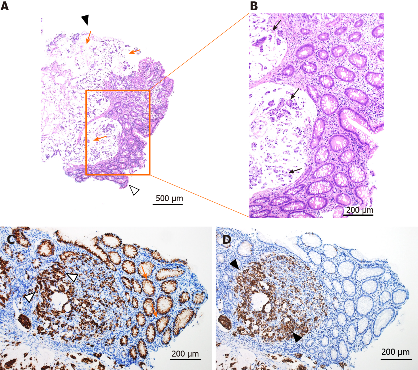
Figure 3 Histological findings of the tumor.
A and B: Hematoxylin and eosin staining. Mucus in the tumor (orange arrows) and adenocarcinoma cells in the mucus (black arrows). Black arrowhead shows the surface of the tumor and white arrowhead shows the bottom of the tumor; C: Mucin 2, oligomeric mucus gel-forming staining (orange arrows and white arrowheads represent positively stained mucosal cells and adenocarcinoma cells); D: Mucin 5AC staining (black arrowheads represent positively stained adenocarcinoma cells).
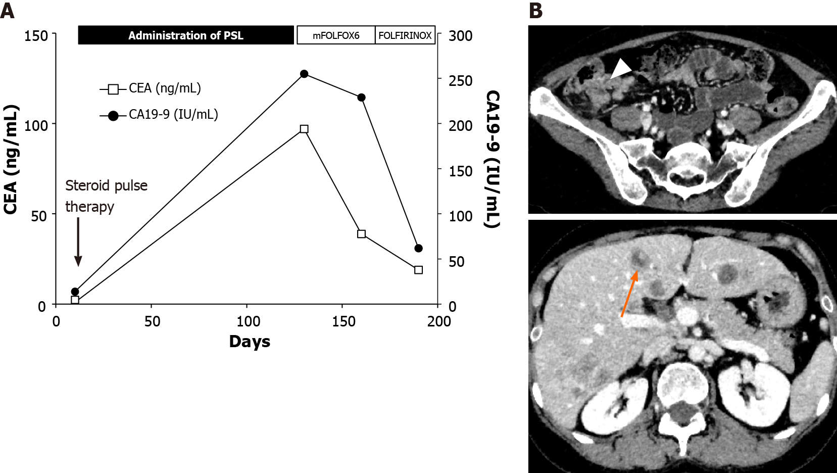
Figure 4 Ultrasonographic and endoscopic images of the tumor.
A: Clinical course of the case; B: Computed tomography results on day 180. CEA: Carcinoembryonic antigen; CA19-9: Carbohydrate antigen 19-9; PSL: Prednisolone; mFOLFOX6: Modified combination of 5-fluorouracil, leucovorin, and oxaliplatin; FOLFIRINOX: Combination of leucovorin, fluorouracil, irinotecan, and oxaliplatin.
- Citation: Koseki Y, Kamimura K, Tanaka Y, Ohkoshi-Yamada M, Zhou Q, Matsumoto Y, Mizusawa T, Sato H, Sakamaki A, Umezu H, Yokoyama J, Terai S. Rapid progression of colonic mucinous adenocarcinoma with immunosuppressive condition: A case report and review of literature. World J Clin Cases 2021; 9(30): 9182-9191
- URL: https://www.wjgnet.com/2307-8960/full/v9/i30/9182.htm
- DOI: https://dx.doi.org/10.12998/wjcc.v9.i30.9182


