©The Author(s) 2021.
World J Clin Cases. Oct 26, 2021; 9(30): 9122-9128
Published online Oct 26, 2021. doi: 10.12998/wjcc.v9.i30.9122
Published online Oct 26, 2021. doi: 10.12998/wjcc.v9.i30.9122
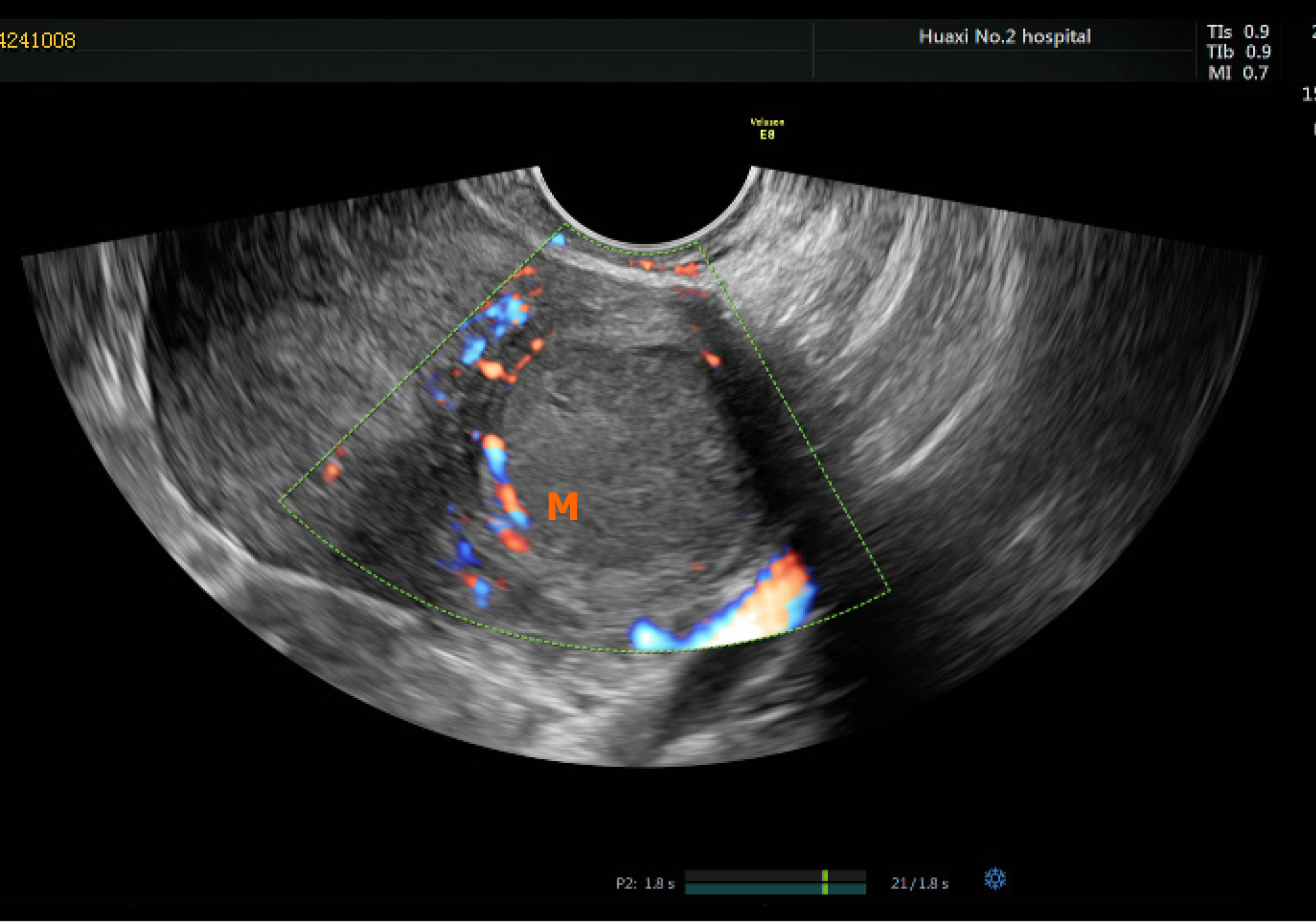
Figure 1 Pelvis transvaginal ultrasonography transverse images showing well-defined isoechoic uterus-like mass.
M: Mass.
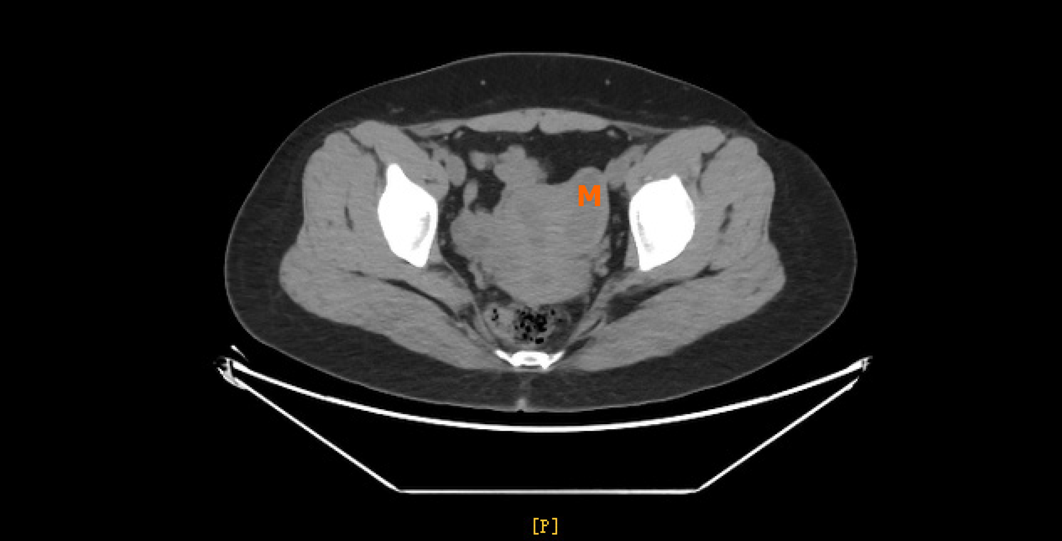
Figure 2 Computed tomography showing a cystic lesion with a size of about 4.
9 cm × 2.0 cm × 2.1 cm on the left side of the pelvis, not clearly demarcated from the left uterine muscle wall. M: Mass.
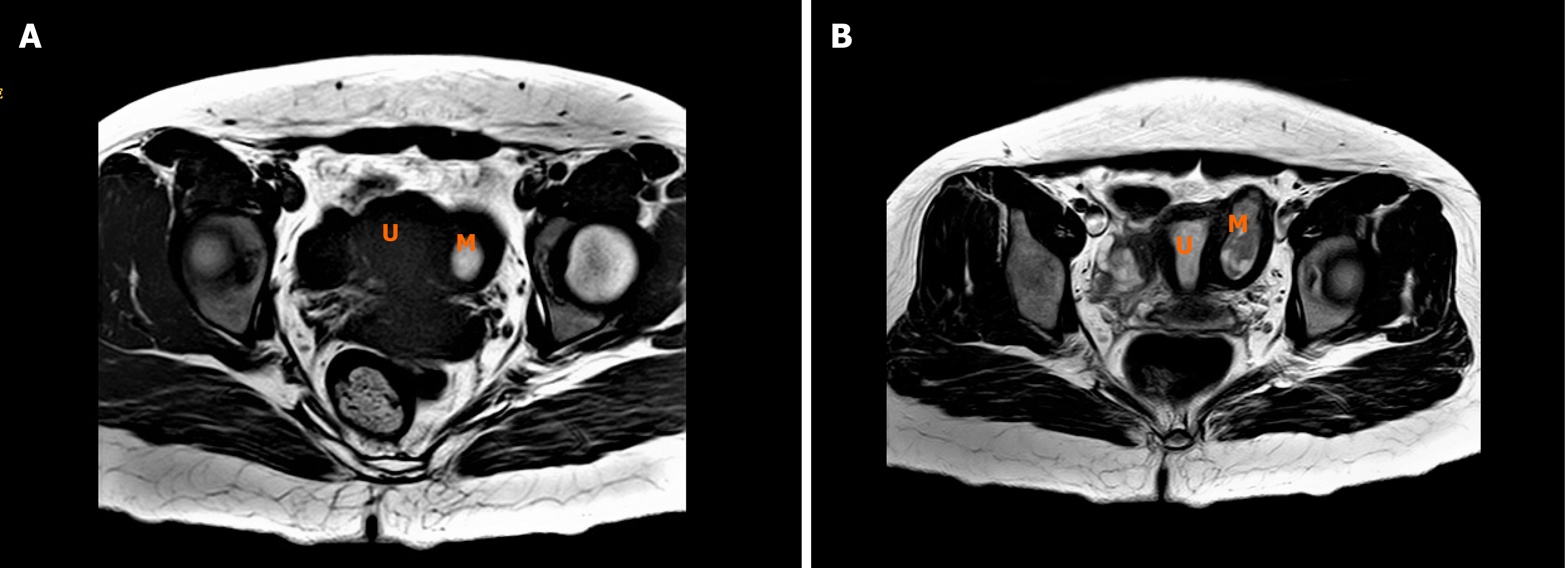
Figure 3 Magnetic resonance imaging showing a cystic lesion with a size of about 5.
0 cm × 2.0 cm × 2.1 cm on the left side wall of the uterus near uterine horn, the cyst fluid signal is uneven, T2W1 showing mixed high signal, T1W1 mainly high signal (A, B). M: Mass; U: Uterus.
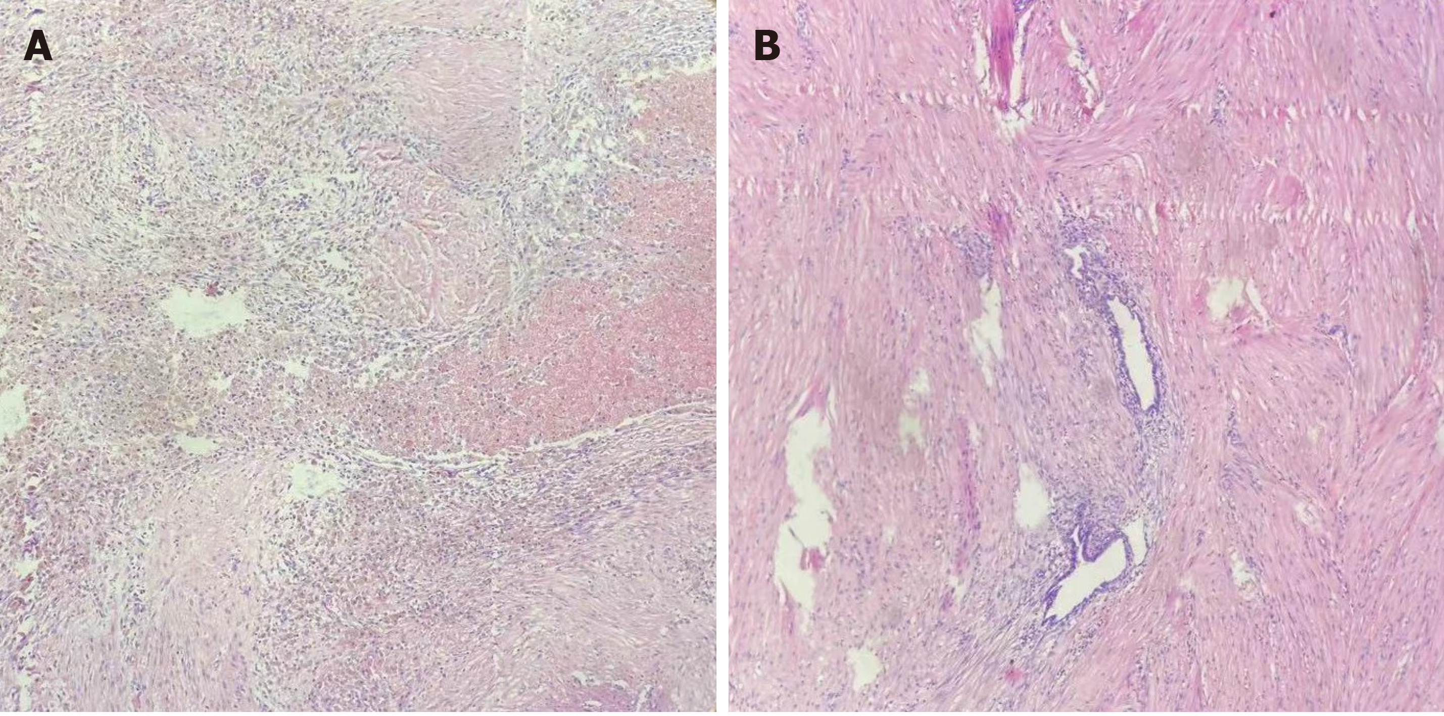
Figure 4 Pathological examination showing functional endometrium, with glands and stroma lining the cavity wall surrounded by irregularly arranged smooth muscle cells, resembling myometrium (200x) (A, B).
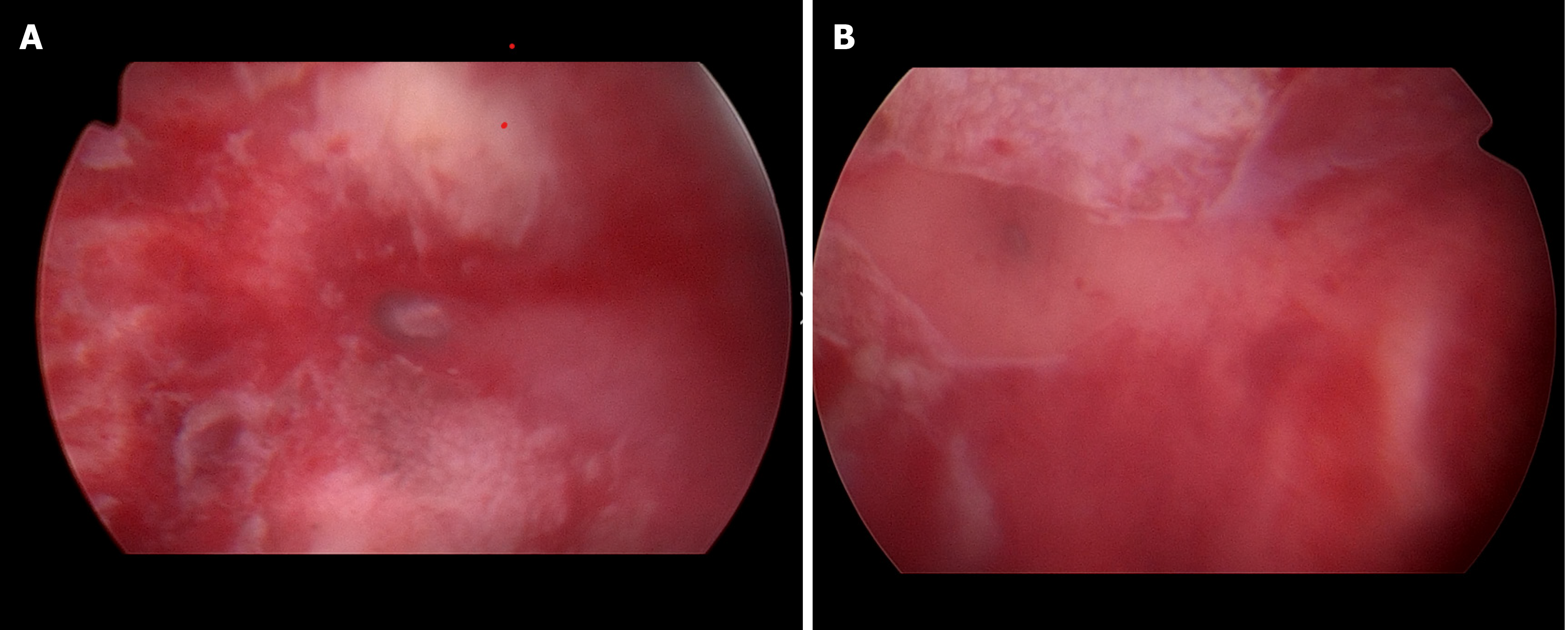
Figure 5 No abnormalities in bilateral cornua uteri and both ostia were visible (A, B).
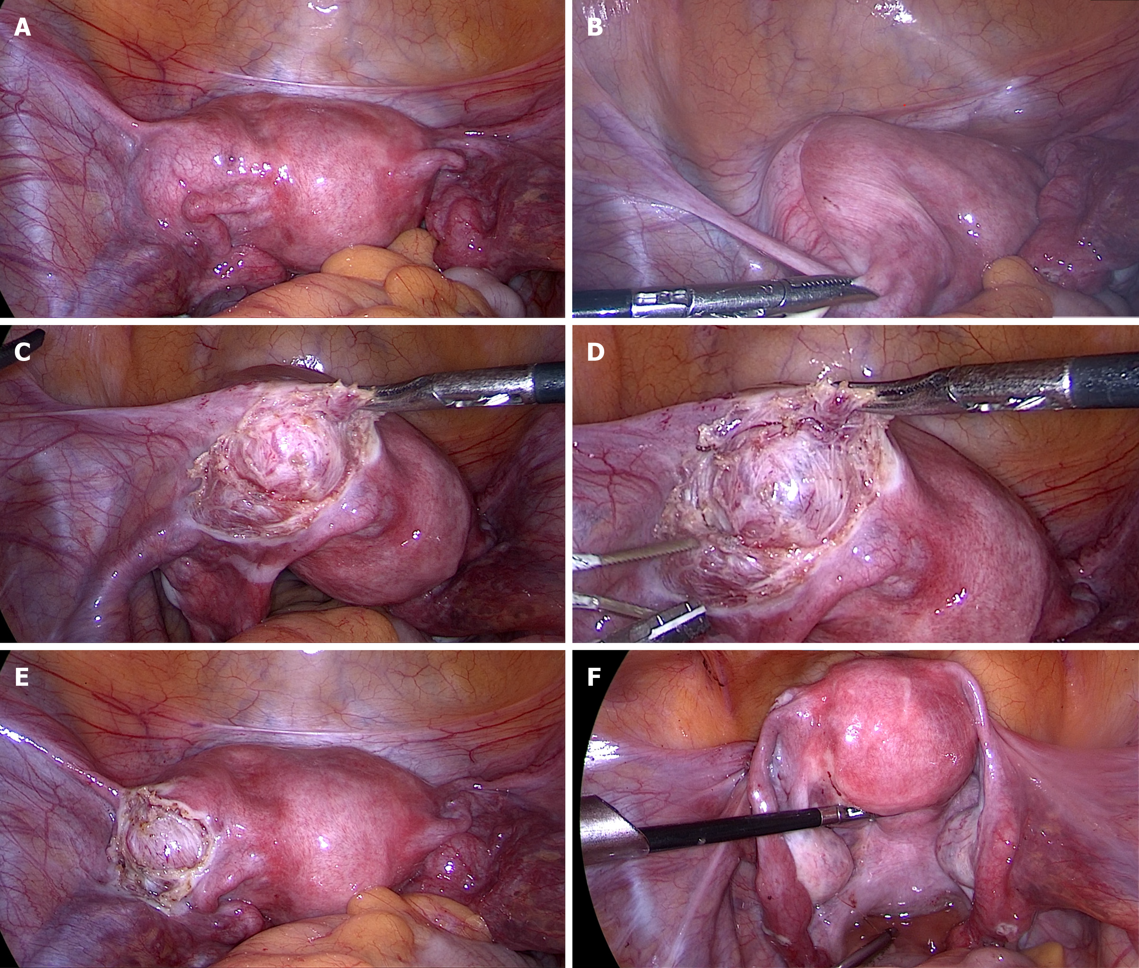
Figure 6 Internal genitalia of the patient showing a left accessory and cavitated uterine mass (A-F).
Uterus showing the accessory and cavitated uterine mass on the left anterior surface. Normal uterus and adnaexae were observed after the exeresis and peritonization.
- Citation: Hu YL, Wang A, Chen J. Diagnosis and laparoscopic excision of accessory cavitated uterine mass in a young woman: A case report. World J Clin Cases 2021; 9(30): 9122-9128
- URL: https://www.wjgnet.com/2307-8960/full/v9/i30/9122.htm
- DOI: https://dx.doi.org/10.12998/wjcc.v9.i30.9122


