Copyright
©The Author(s) 2021.
World J Clin Cases. Oct 16, 2021; 9(29): 8888-8893
Published online Oct 16, 2021. doi: 10.12998/wjcc.v9.i29.8888
Published online Oct 16, 2021. doi: 10.12998/wjcc.v9.i29.8888
Figure 1 Preoperative and postoperative imaging.
A: Computed tomography (CT) revealed a mass occluding about 90% of the tracheal lumen; B: Contrast-enhanced CT a soft tissue mass with obvious enhancement on the right posterolateral wall of the trachea; C: CT indicated a slight protrusion at the position of the incision.
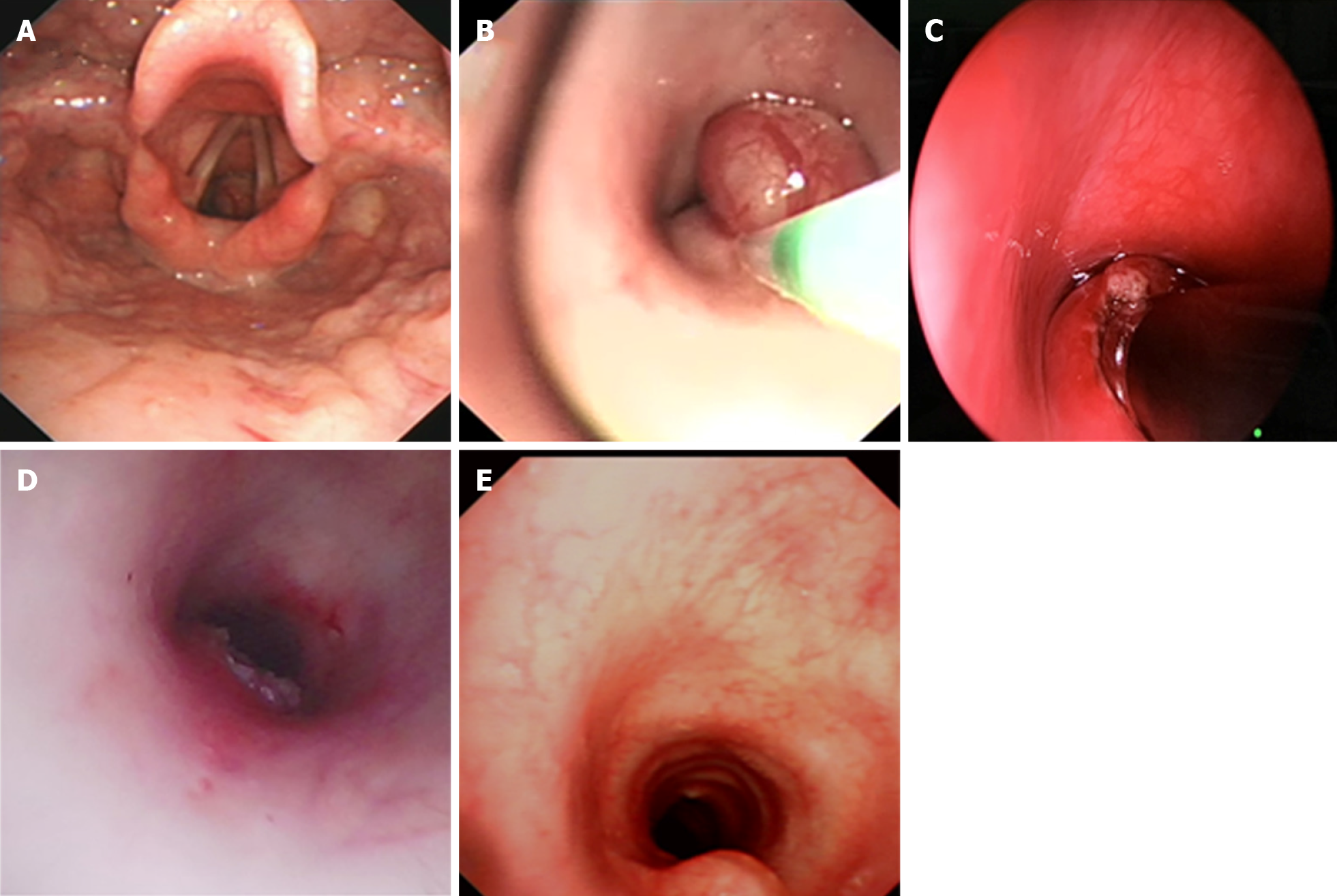
Figure 2 Fiberoptic bronchoscopy findings.
A: A mass was located in the trachea at the 4- to 7-o’clock position about 1 cm below the vocal cords; B: The spherical part of the mass was removed by an electrocautery snare; C: The base was ablated by a low-temperature ion probe; D: Most of the base was removed; E: There was a slight protrusion at the position of the incision.
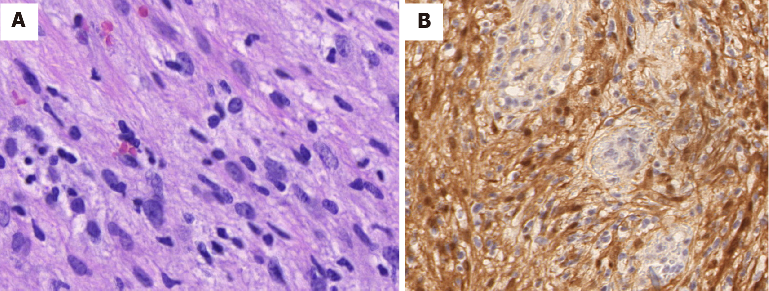
Figure 3 Pathological images of tumor cells.
A: A histologic section from the tumor shows that the tumor cells were spindled with elongated palisading nuclei (hematoxylin–eosin stain, 400 ×); B: The tumor cells showed diffuse and positive S-100 staining by immunohistochemistry (streptavidin peroxidase method, 200 ×).
- Citation: Wu L, Sha MC, Wu XL, Bi J, Chen ZM, Wang YS. Primary intratracheal neurilemmoma in a 10-year-old girl: A case report. World J Clin Cases 2021; 9(29): 8888-8893
- URL: https://www.wjgnet.com/2307-8960/full/v9/i29/8888.htm
- DOI: https://dx.doi.org/10.12998/wjcc.v9.i29.8888


