©The Author(s) 2021.
World J Clin Cases. Oct 16, 2021; 9(29): 8879-8887
Published online Oct 16, 2021. doi: 10.12998/wjcc.v9.i29.8879
Published online Oct 16, 2021. doi: 10.12998/wjcc.v9.i29.8879
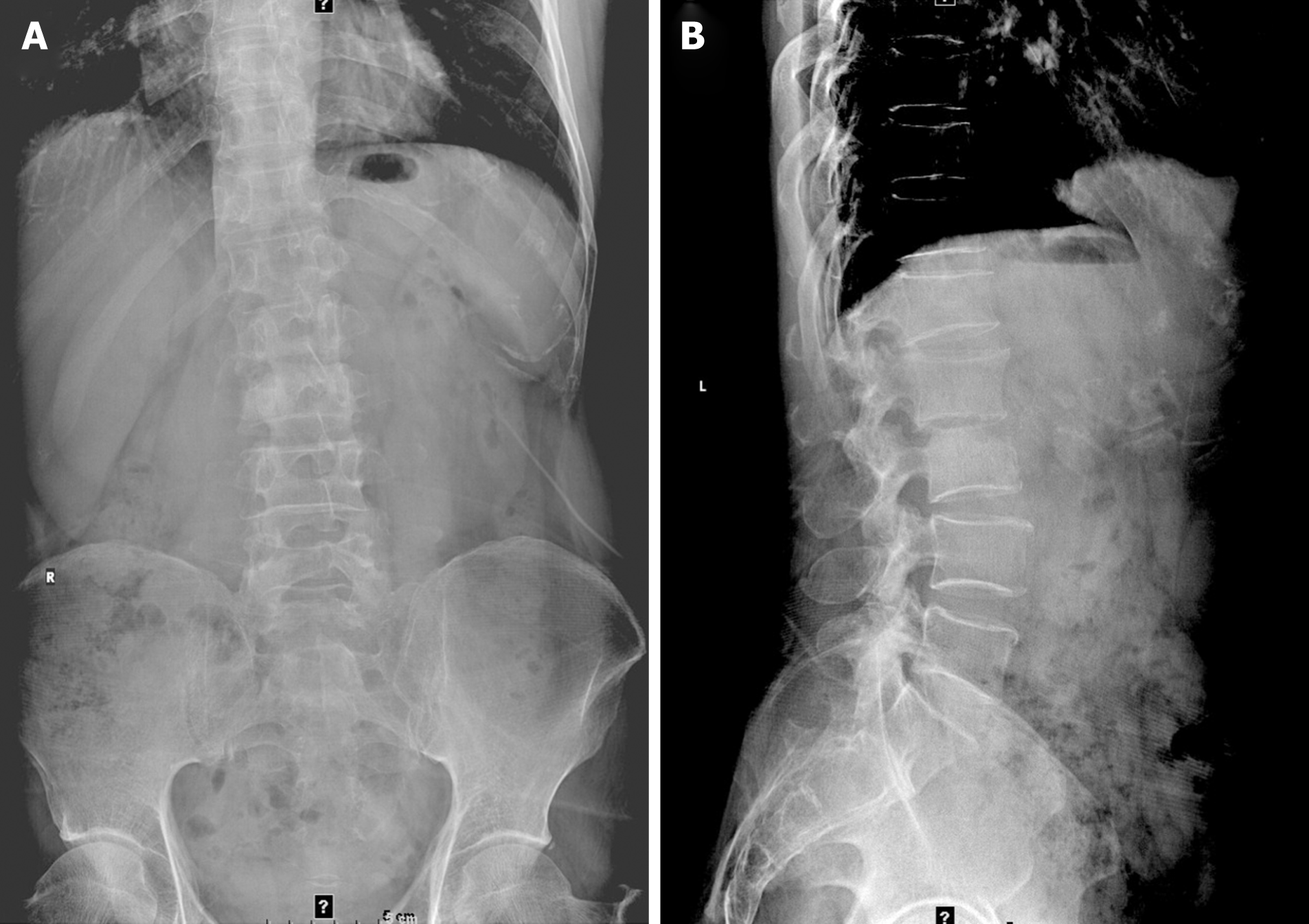
Figure 1 Lumbar spine X-ray showing L1 and L2 vertebral body localized bone destruction with a narrowing of this vertebral space.
A: Anteroposterior radiograph of the lumbar spine; B: Lateral radiograph of the lumbar spine.
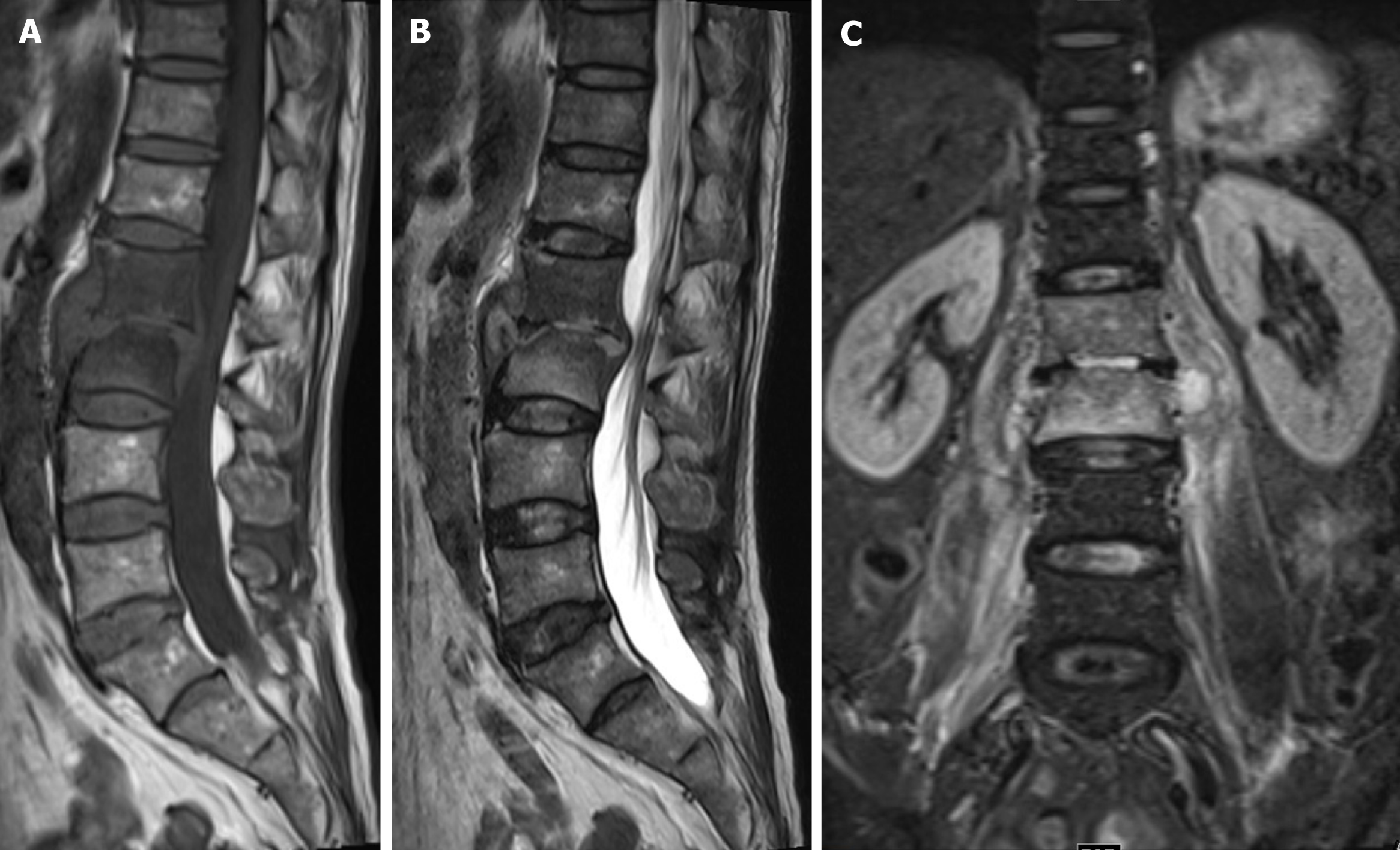
Figure 2 Magnetic resonance imaging of the lumbar spine showing bone destruction of the L1 and L2 vertebrae, a narrowing of the L1/2 intervertebral space, the destruction of the intervertebral disc, and obvious swelling of the surrounding soft tissues.
A: T1-magnetic resonance imaging (MRI) imaging; B: T2-weighted MRI imaging; C: Lumbar coronal MRI image.
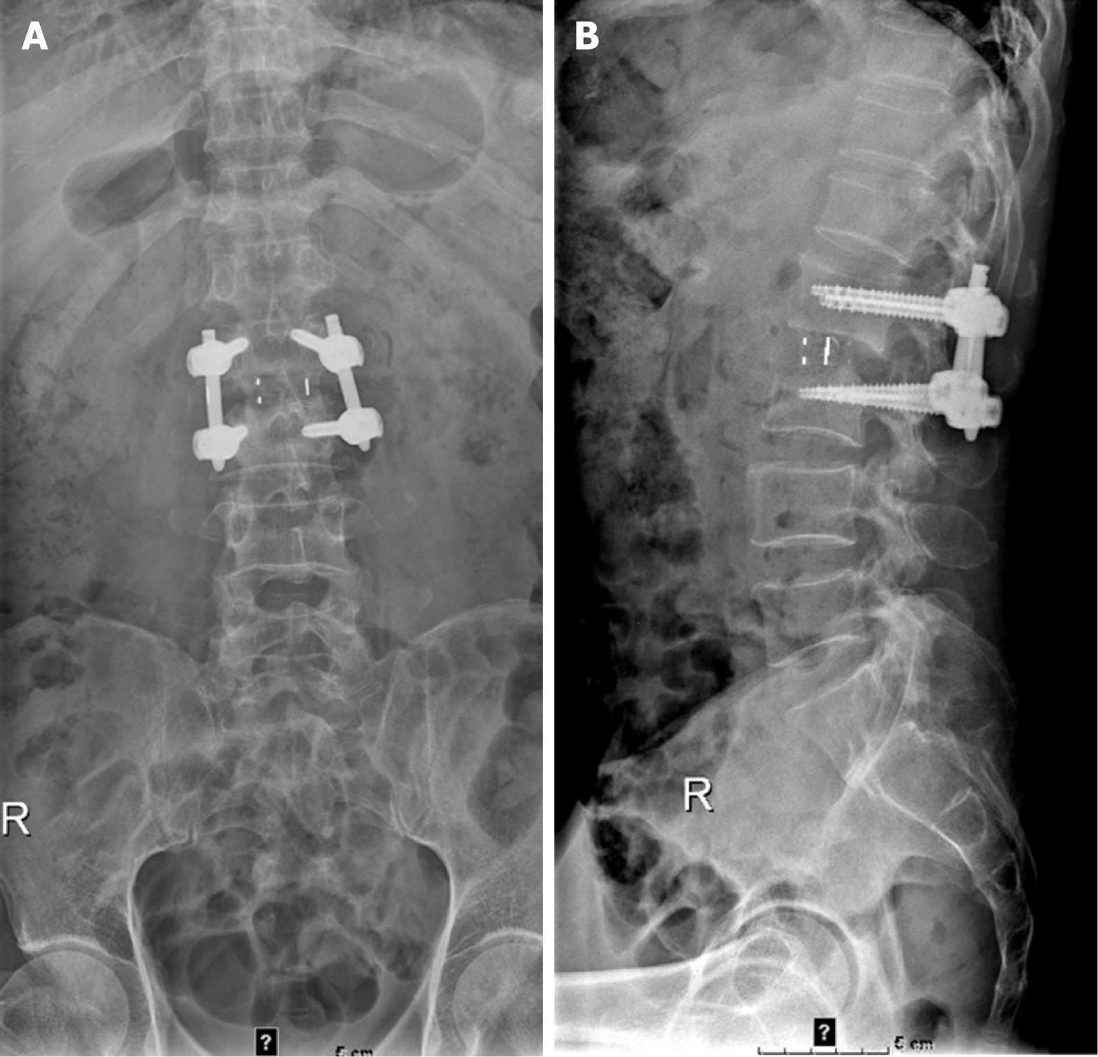
Figure 3 Lumbar spine X-rays showing that the L1-L2 interbody internal fixation device was not broken, dislodged, or displaced, and the intervertebral bone graft had healed well.
A: Anteroposterior radiograph of the lumbar spine; B: Lateral radiograph of the lumbar spine.
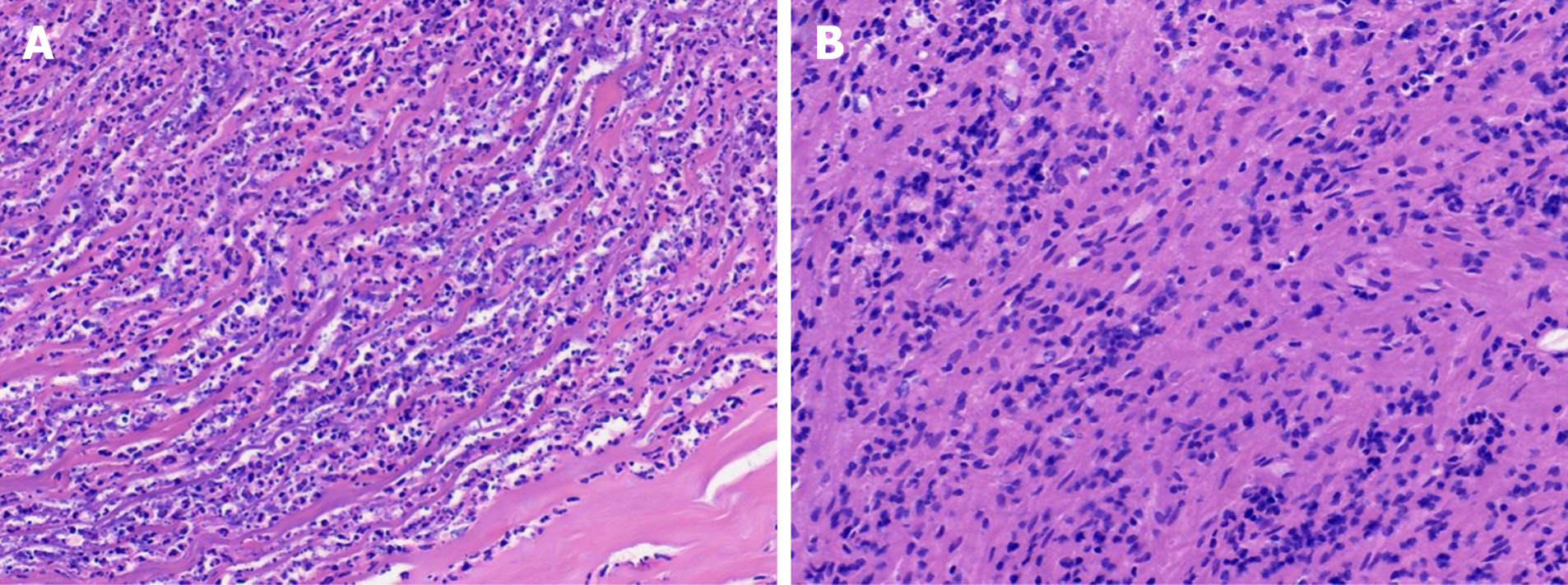
Figure 4 Histological staining of lumbar 1/2 focal tissue.
A: Haematoxylin-eosin staining (40 × magnification) showing acute purulent inflammation and inflammatory necrosis; B: Haematoxylin-eosin staining (magnification × 40) showing chronic inflammatory cell infiltration.
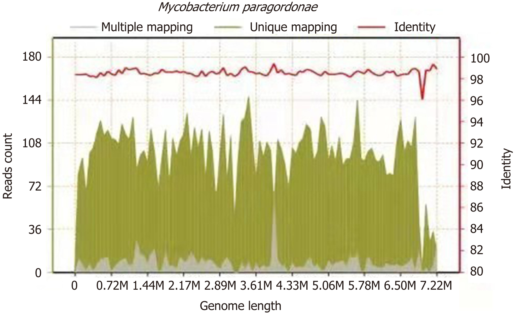
Figure 5 Metagenomic next-generation sequencing suggesting the presence of Mycobacterium paragordonae.
The total number of bases in the genome of this species was 7224251, the total length of sequence coverage was 618365 bp, the coverage degree was 8.559572%, and the average depth was 1.30x.
- Citation: Tan YZ, Yuan T, Tan L, Tian YQ, Long YZ. Lumbar infection caused by Mycobacterium paragordonae: A case report. World J Clin Cases 2021; 9(29): 8879-8887
- URL: https://www.wjgnet.com/2307-8960/full/v9/i29/8879.htm
- DOI: https://dx.doi.org/10.12998/wjcc.v9.i29.8879


