Copyright
©The Author(s) 2021.
World J Clin Cases. Oct 6, 2021; 9(28): 8509-8517
Published online Oct 6, 2021. doi: 10.12998/wjcc.v9.i28.8509
Published online Oct 6, 2021. doi: 10.12998/wjcc.v9.i28.8509
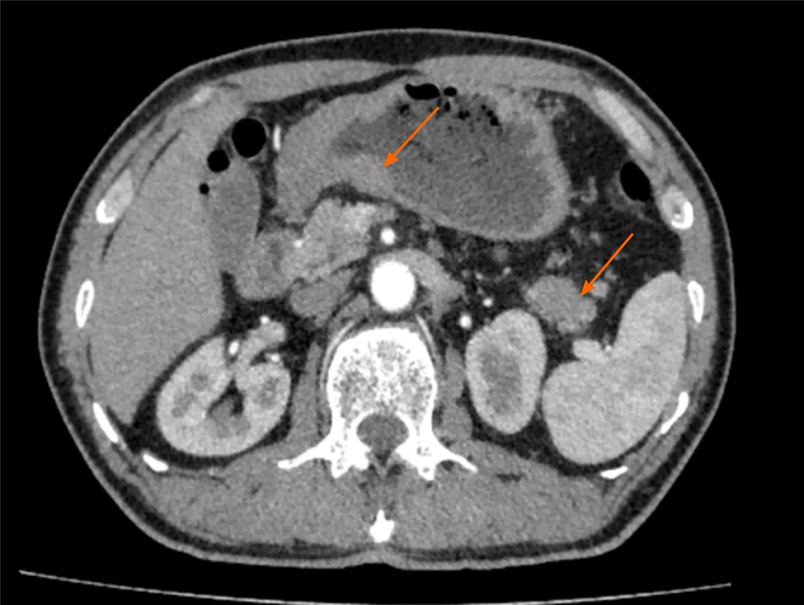
Figure 1 Imaging examinations performed before surgery.
On contrast-enhanced computed tomography of stomach, arrow on the left showed uneven thickened with irregular mucosa and heterogeneous contrast enhancement on the antrum of gastric wall; arrow on the right indicated a space-occupying lesion about 34 mm × 16 mm in the tail of the pancreas.
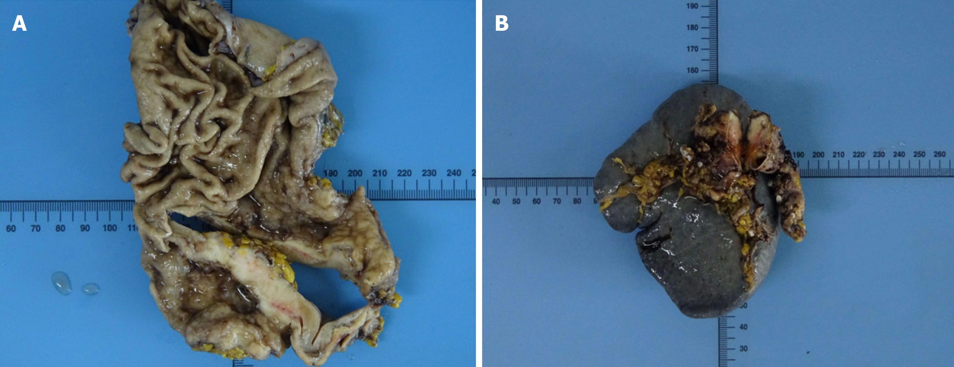
Figure 2 Resection specimen.
A: Resection specimen of gastric tumor; B: Resection specimen of pancreatic tumor.
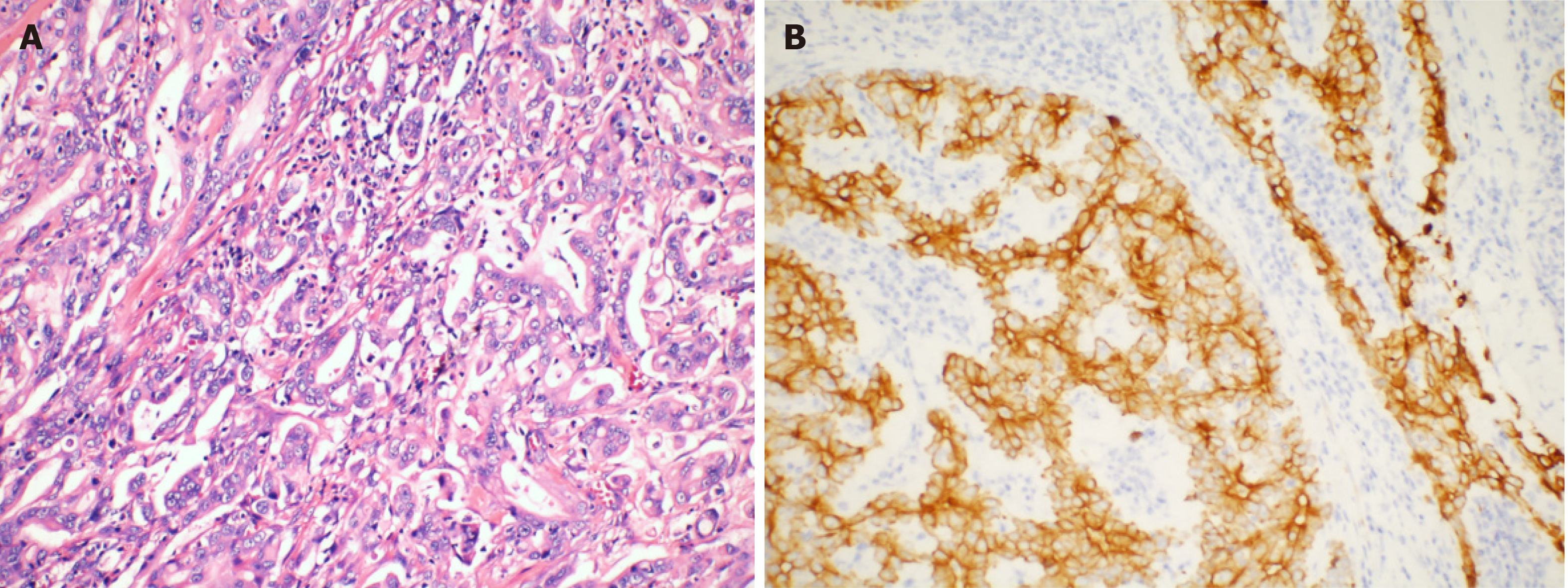
Figure 3 Microscopic examinations of gastric.
A: Routine histology, stained using hematoxylin-eosin, shows gastric adenocarcinoma (magnification: × 200); B: Immunohistochemical staining of gastric tumor cells is partial positive for Cytokeratin 7 (magnification: × 200).
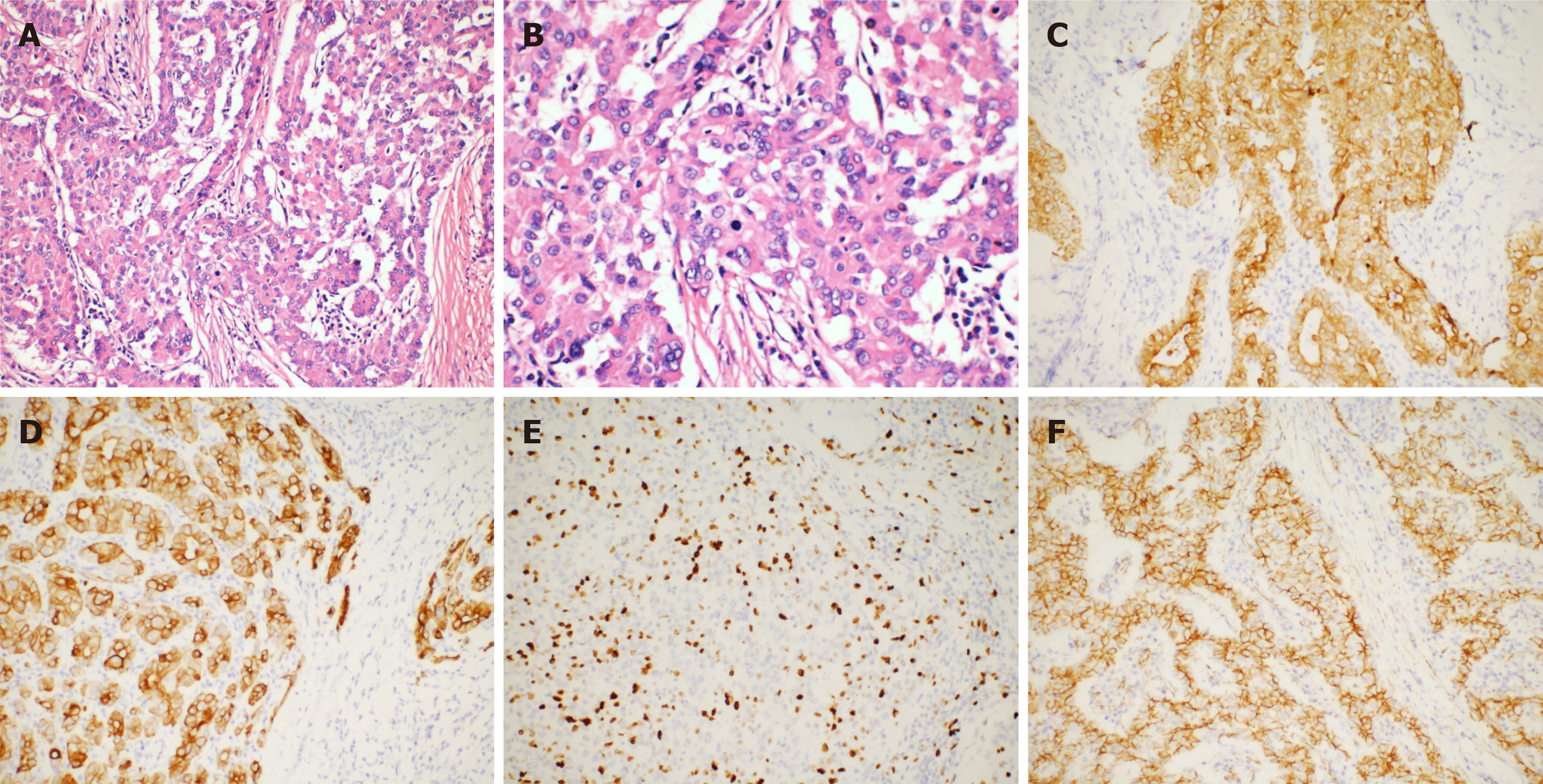
Figure 4 Microscopic examinations of pancreas.
A: Routine histology, stained using hematoxylin-eosin, shows pancreatic acinar cell adenocarcinoma (magnification: × 200); B: Nuclear division in pancreatic acinar cell carcinoma (magnification: × 400); C: Immunohistochemical staining of pancreatic tumor cells: CAM5.2 expression in pancreatic tumor (magnification: × 200); D: CK19 expression in pancreatic tumor (magnification: × 200); E: Ki-67 partial expression in pancreatic tumor (+ 30%) (magnification: × 200); F: Membranous expression of beta-catenin in pancreatic tumor (magnification: × 200).
- Citation: Fang T, Liang TT, Wang YZ, Wu HT, Liu SH, Wang C. Synchronous concomitant pancreatic acinar cell carcin and gastric adenocarcinoma: A case report and review of literature. World J Clin Cases 2021; 9(28): 8509-8517
- URL: https://www.wjgnet.com/2307-8960/full/v9/i28/8509.htm
- DOI: https://dx.doi.org/10.12998/wjcc.v9.i28.8509


