Copyright
©The Author(s) 2021.
World J Clin Cases. Sep 26, 2021; 9(27): 8214-8219
Published online Sep 26, 2021. doi: 10.12998/wjcc.v9.i27.8214
Published online Sep 26, 2021. doi: 10.12998/wjcc.v9.i27.8214
Figure 1 Perioperative images.
A: Perioperative computed tomography (CT) angiogram of the female patient; B and C: Perioperative magnetic resonance cholangiopancreatography and CT images of the male patient.
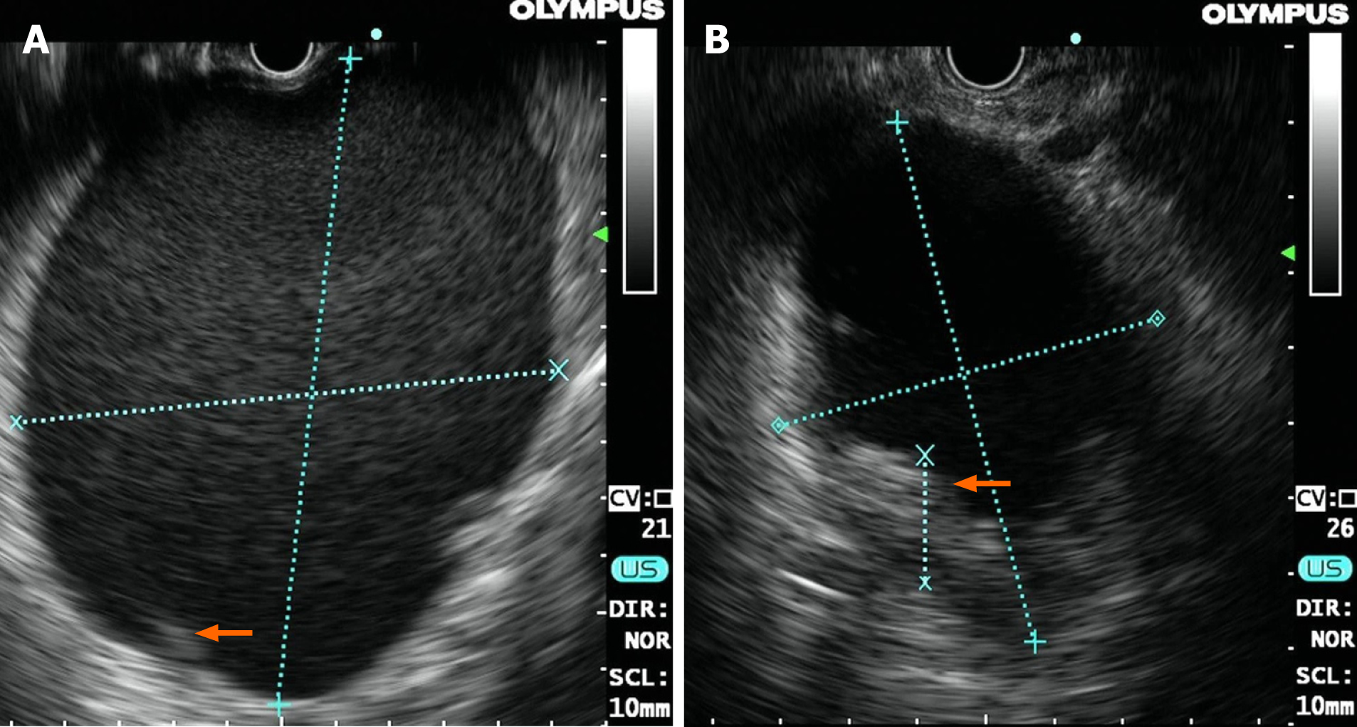
Figure 2 Endoscopic ultrasound images of the lesion in each patient.
A: The size and high-density shadows (arrow) of the lesion in the female on endoscopic ultrasound (EUS); B: The size and high-density shadows (arrow) of the lesion in the male on EUS.
Figure 3 Direct observation of multiple hidden vessels under endoscopy.
A: Multiple hidden vessels in the female; B: Multiple hidden vessels in the male.
Figure 4 Retrospective observation of computed tomography images.
A: Radiological signs of nonliquid contents and some cord-like shadows (arrow) in the female; B: Solid debris (arrow) in the male.
- Citation: Xu N, Zhai YQ, Li LS, Chai NL. Multiple hidden vessels in walled-off necrosis with high-risk bleeding: Report of two cases. World J Clin Cases 2021; 9(27): 8214-8219
- URL: https://www.wjgnet.com/2307-8960/full/v9/i27/8214.htm
- DOI: https://dx.doi.org/10.12998/wjcc.v9.i27.8214


