Copyright
©The Author(s) 2021.
World J Clin Cases. Sep 26, 2021; 9(27): 8199-8206
Published online Sep 26, 2021. doi: 10.12998/wjcc.v9.i27.8199
Published online Sep 26, 2021. doi: 10.12998/wjcc.v9.i27.8199
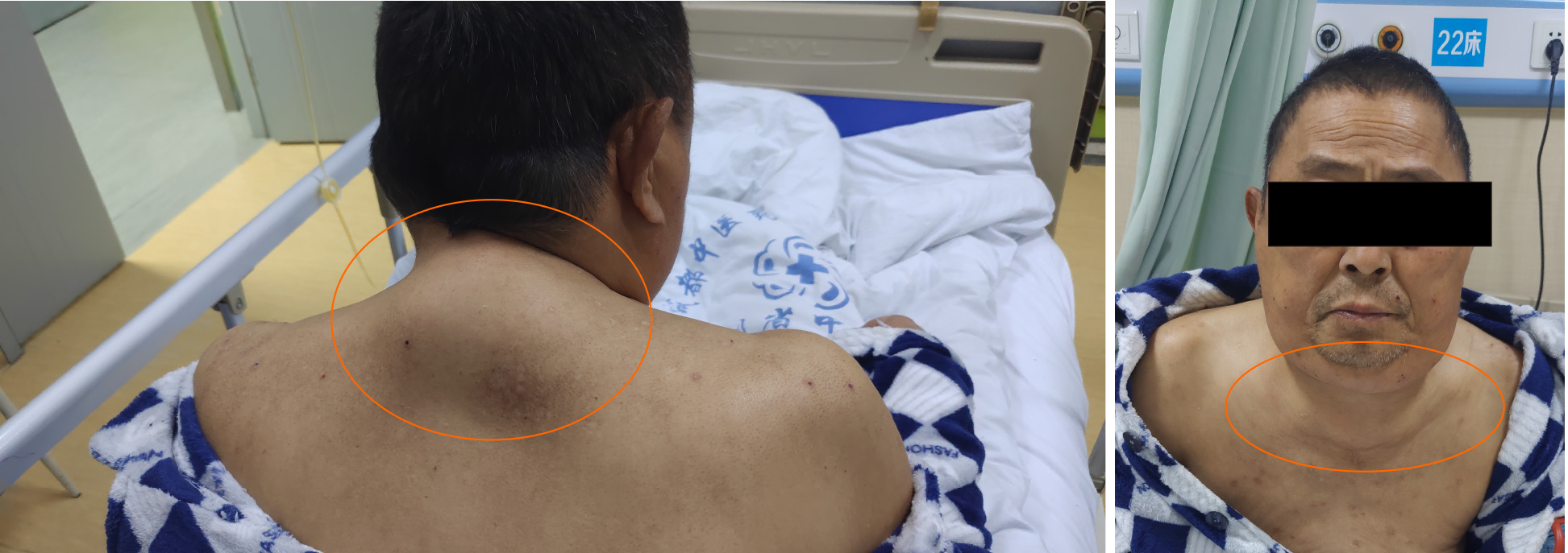
Figure 1 The patient at presentation.
Symmetrical lumps were obvious around the neck, featuring normal skin color and no ulceration or exudation.
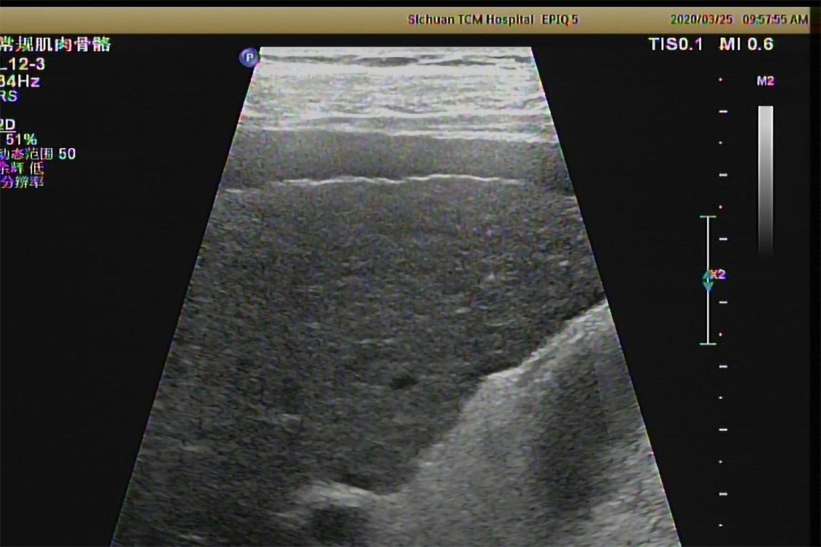
Figure 2 Ultrasound image of the patient’s liver.
Chronic liver damage is seen.
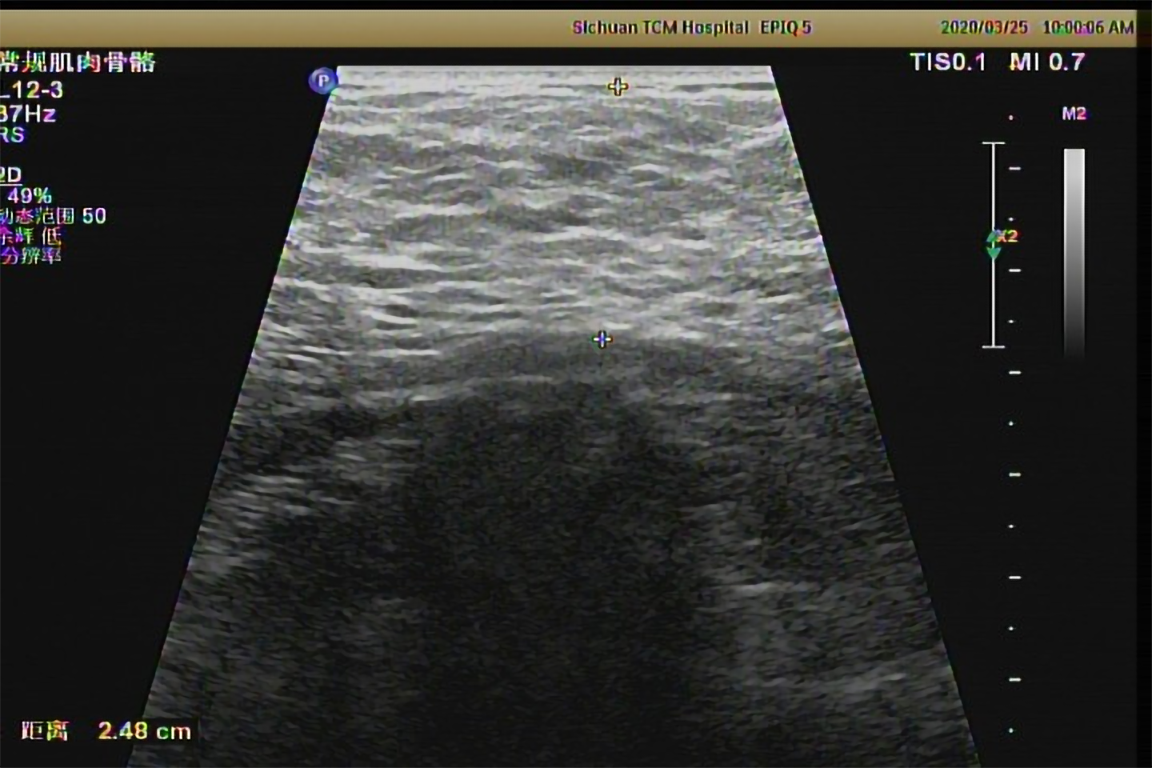
Figure 3 Ultrasound image of a neck mass in the patient.
The subcutaneous fatty masses are seen in the cervical-supraclavicular and occipital regions, being significantly enhanced on both sides, with the thickness of 2.48 cm and unclear borders.
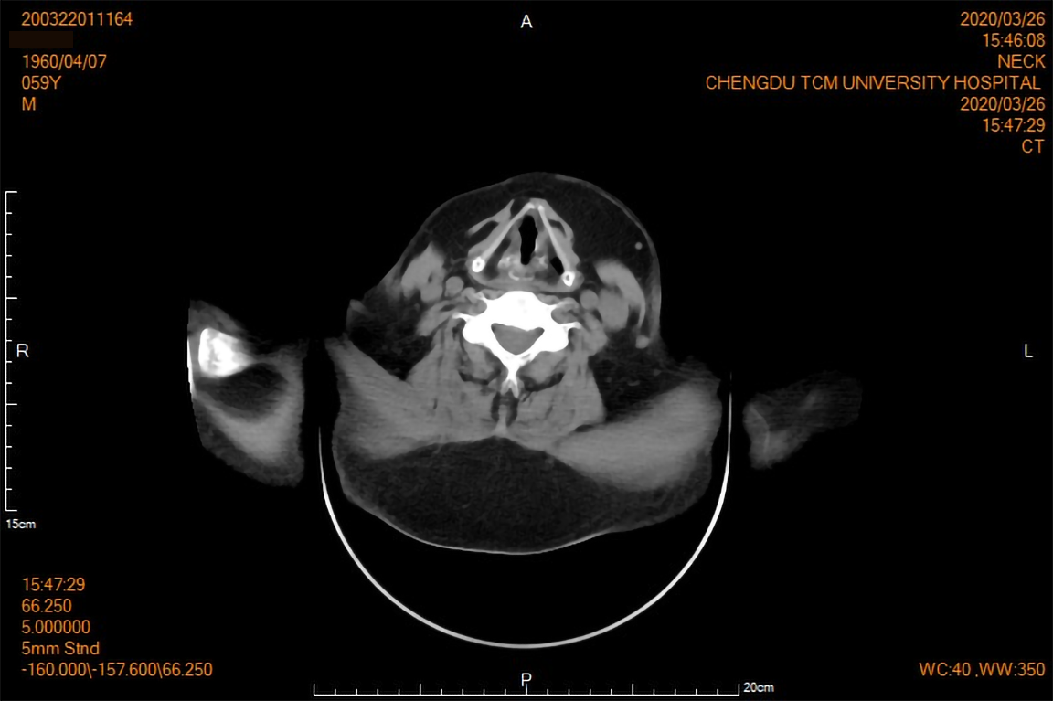
Figure 4 Computed tomography image showing the subcutaneous fat layer around the neck (including the submandibular region) to be significantly thickened and symmetrically distributed on both sides.
Morphology of the oropharynx and laryngo-pharynx was normal. No obvious stenosis was observed.
- Citation: Wu L, Jiang T, Zhang Y, Tang AQ, Wu LH, Liu Y, Li MQ, Zhao LB. Madelung’s disease with alcoholic liver disease and acute kidney injury: A case report. World J Clin Cases 2021; 9(27): 8199-8206
- URL: https://www.wjgnet.com/2307-8960/full/v9/i27/8199.htm
- DOI: https://dx.doi.org/10.12998/wjcc.v9.i27.8199


