Copyright
©The Author(s) 2021.
World J Clin Cases. Sep 26, 2021; 9(27): 8171-8176
Published online Sep 26, 2021. doi: 10.12998/wjcc.v9.i27.8171
Published online Sep 26, 2021. doi: 10.12998/wjcc.v9.i27.8171
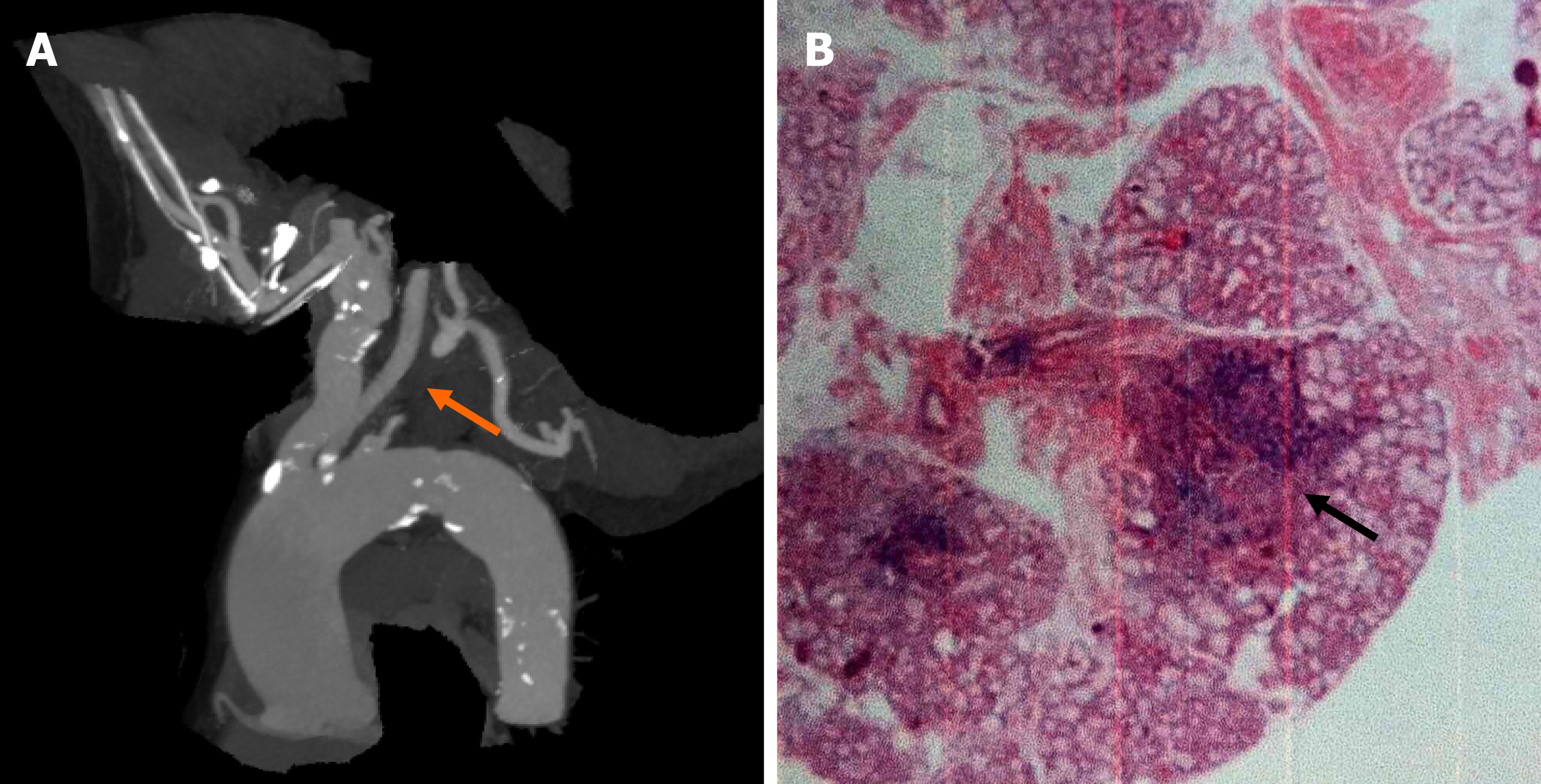
Figure 1 Results of labial gland biopsy and aortic angiography.
A: Aortic angiography showing that the proximal part of the left subclavian artery was occluded, and a small ulcer of the aortic arch was formed; B: Salivary gland tissue showing chronic inflammation, including five lymphatic foci.
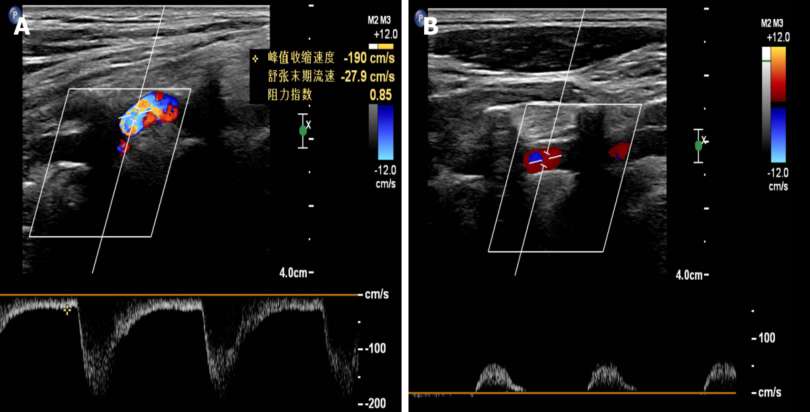
Figure 2 Color Doppler ultrasound of the vertebral arteries.
A: Blood flow velocity of the right vertebral artery was 190 cm/s, with a resistance index of 0.85; B: The blood flow of the left vertebral artery was reversed, showing "small slow waves".
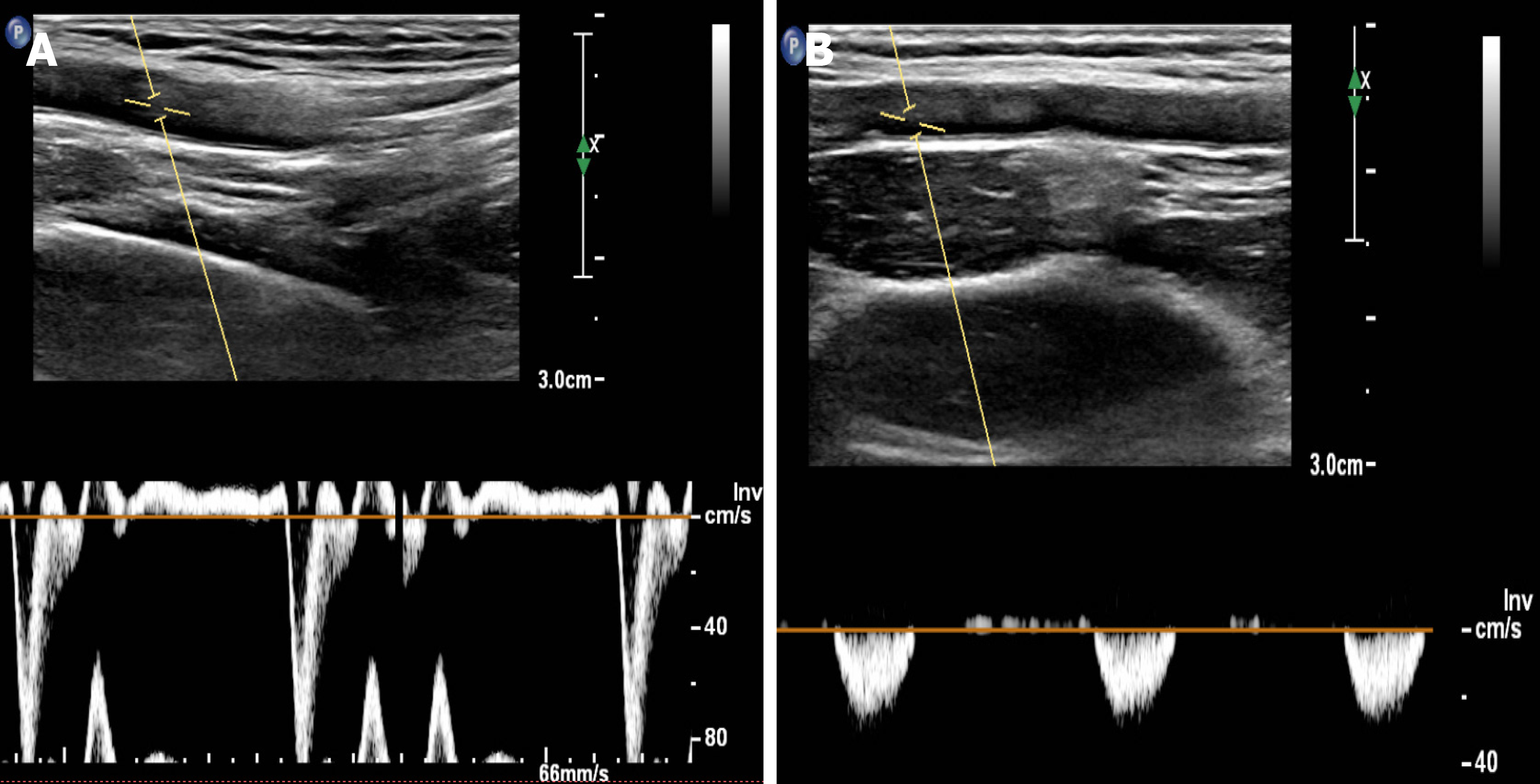
Figure 3 Color Doppler ultrasound of the upper extremity arteries.
A: The right upper extremity artery is normal; B: The frequency spectrum of the left upper extremity artery is a "small slow-wave".
- Citation: Hao LJ, Zhang J, Naveed M, Chen KY, Xiao PX. Subclavian steal syndrome associated with Sjogren's syndrome: A case report. World J Clin Cases 2021; 9(27): 8171-8176
- URL: https://www.wjgnet.com/2307-8960/full/v9/i27/8171.htm
- DOI: https://dx.doi.org/10.12998/wjcc.v9.i27.8171


