Copyright
©The Author(s) 2021.
World J Clin Cases. Sep 26, 2021; 9(27): 8127-8134
Published online Sep 26, 2021. doi: 10.12998/wjcc.v9.i27.8127
Published online Sep 26, 2021. doi: 10.12998/wjcc.v9.i27.8127
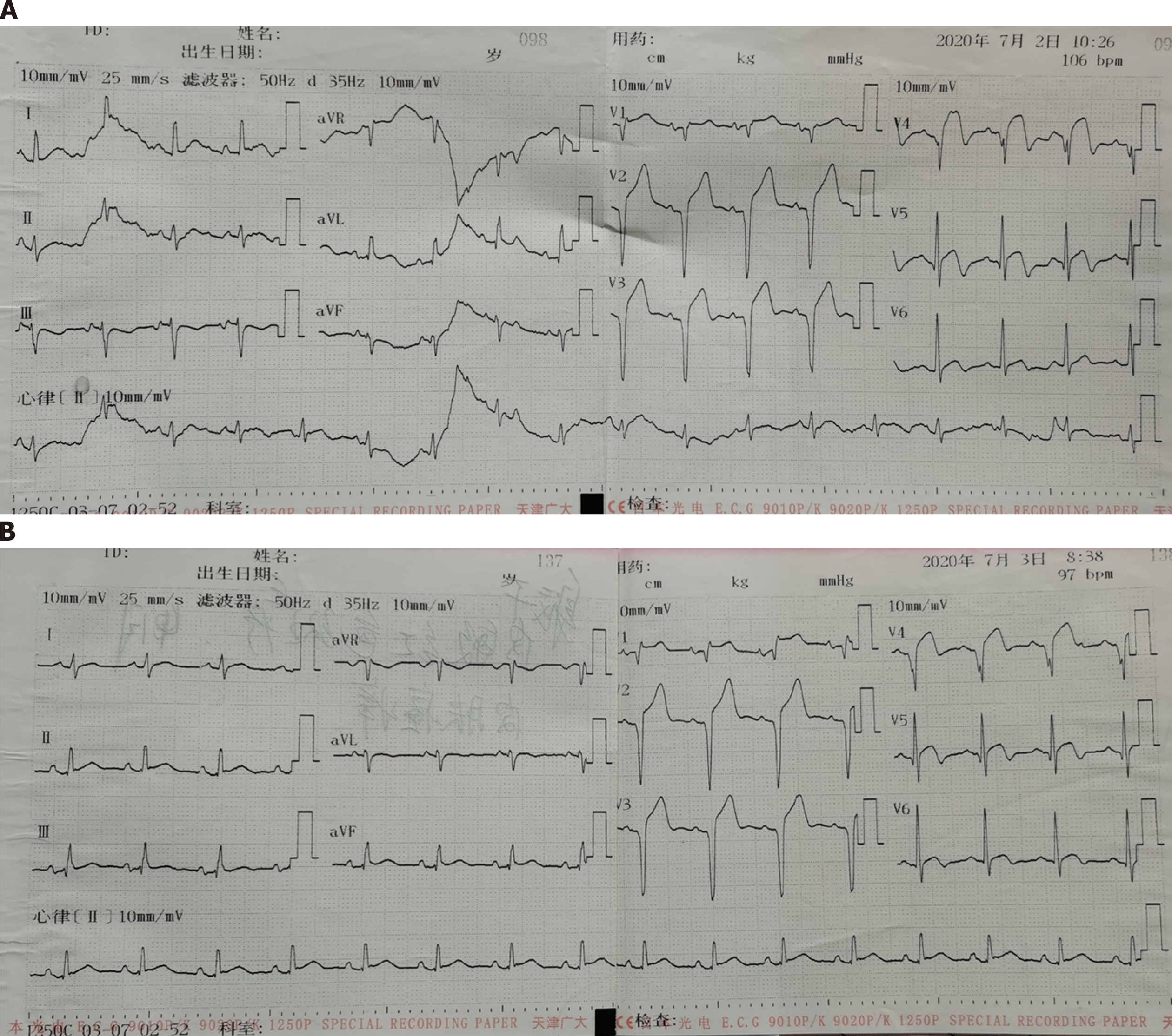
Figure 1 Electrocardiogram examination.
A: On admission; B: Day after admission.
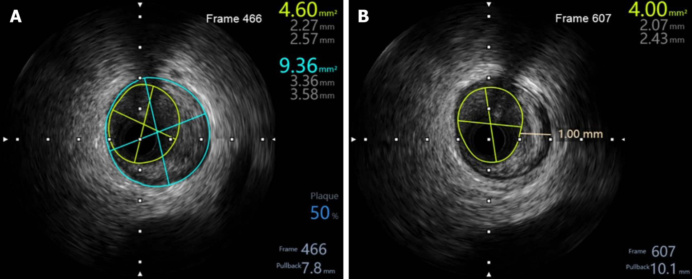
Figure 2 Presence of plaque in the middle of the anterior descending coronary artery.
A: Plaque burden was 50%; B: Plaque thickness was 1 mm.
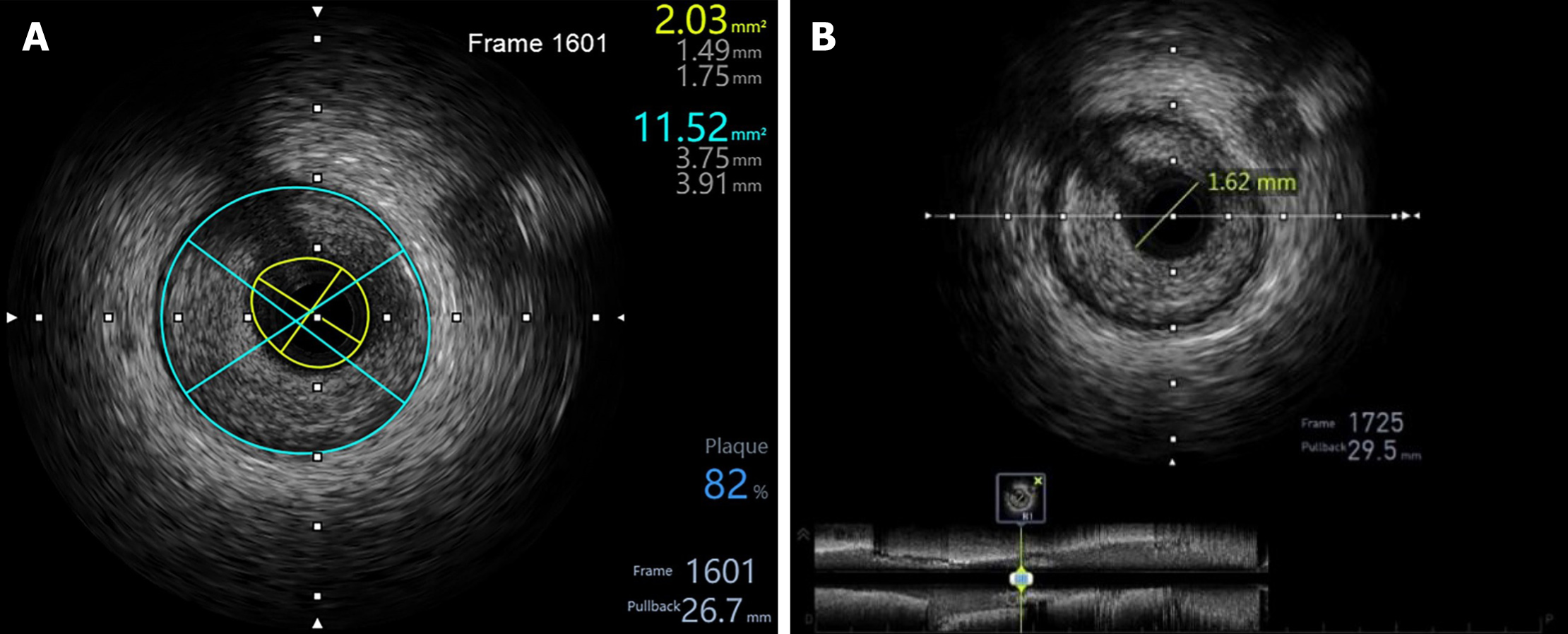
Figure 3 Plaque features before interventional treatment of the proximal anterior descending coronary artery.
A: Plaque burden was 82%, with minimum lumen cross-sectional area of 2.03 mm2; B: Lumen diameter was 1.62 mm.
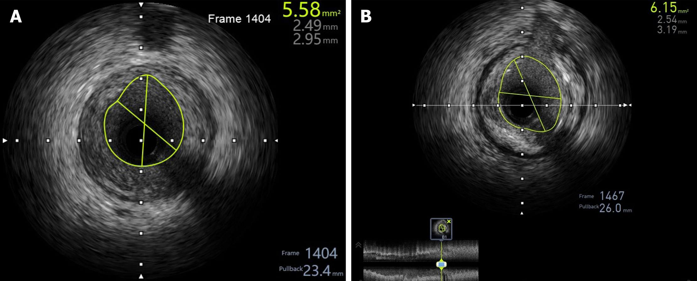
Figure 4 Plaque features after interventional treatment of the proximal anterior descending coronary artery.
A: Lumen cross-sectional area (CSA) was 5.58 mm2; B: Maximum lumen CSA was 6.15 mm2.
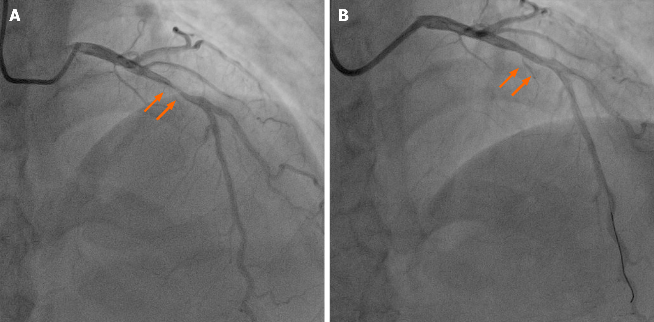
Figure 5 Coronary angiography of the proximal anterior descending coronary artery.
A: Before interventional treatment, stenosis was 95%; B: After interventional treatment, the stenosis obviously relieved. Orange arrows indicate the narrowest position.
- Citation: Zhu HM, Zhang Y, Tang Y, Yuan H, Li ZX, Long Y. Acute coronary syndrome with severe atherosclerotic and hyperthyroidism: A case report. World J Clin Cases 2021; 9(27): 8127-8134
- URL: https://www.wjgnet.com/2307-8960/full/v9/i27/8127.htm
- DOI: https://dx.doi.org/10.12998/wjcc.v9.i27.8127


