Copyright
©The Author(s) 2021.
World J Clin Cases. Sep 26, 2021; 9(27): 8104-8113
Published online Sep 26, 2021. doi: 10.12998/wjcc.v9.i27.8104
Published online Sep 26, 2021. doi: 10.12998/wjcc.v9.i27.8104
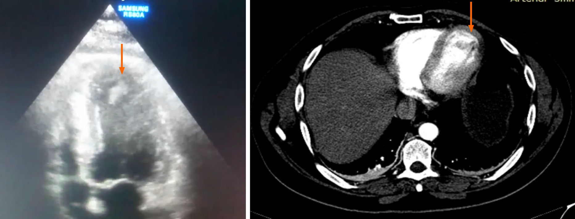
Figure 1 Transthoracic echocardiography.
Hypertrophied left ventricle and hyperechogenic mass at the apex consistent with tongue-shaped mobile thrombus was confirmed by the thoracic computed tomography scan.
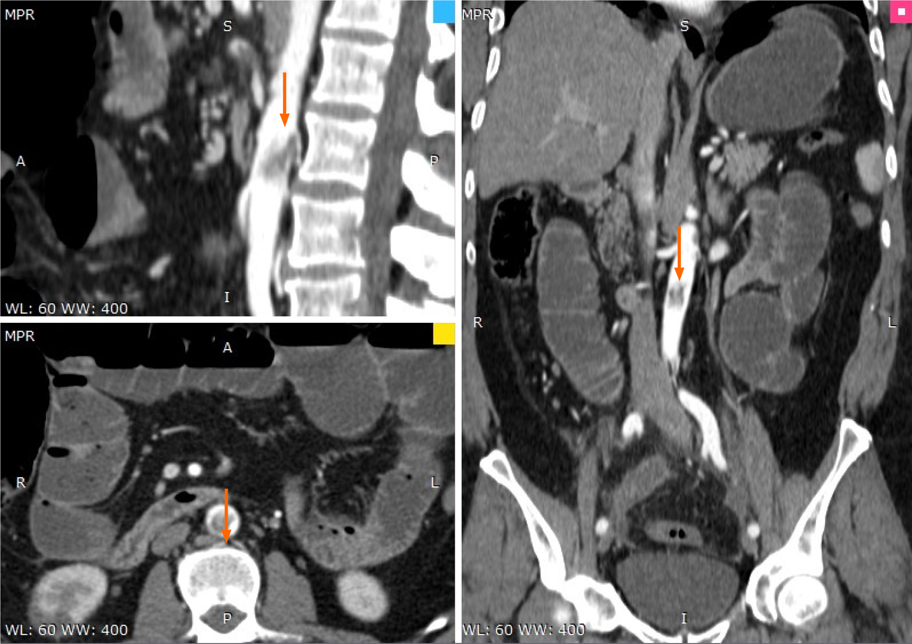
Figure 2 Computed tomography abdominal scan – subocclusion of the superior mesenteric artery.
Figure 3 Computed tomography thoraco-abdominal scan – thrombosis of the posterior aortic wall.
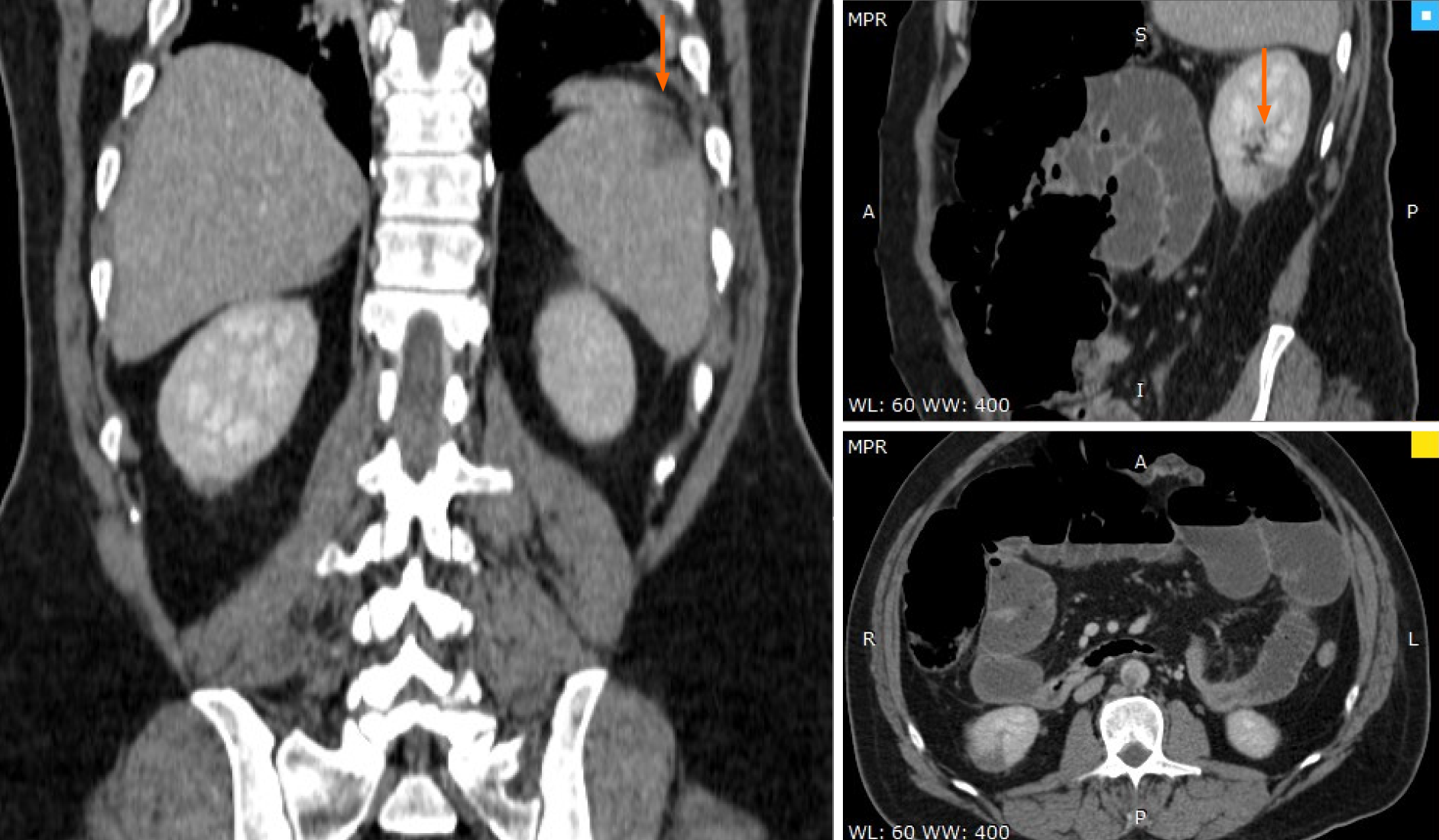
Figure 4 Computed tomography abdominal scan – spleen (superior pole) and right renal infarction (inferior pole) and ileum necrosis.
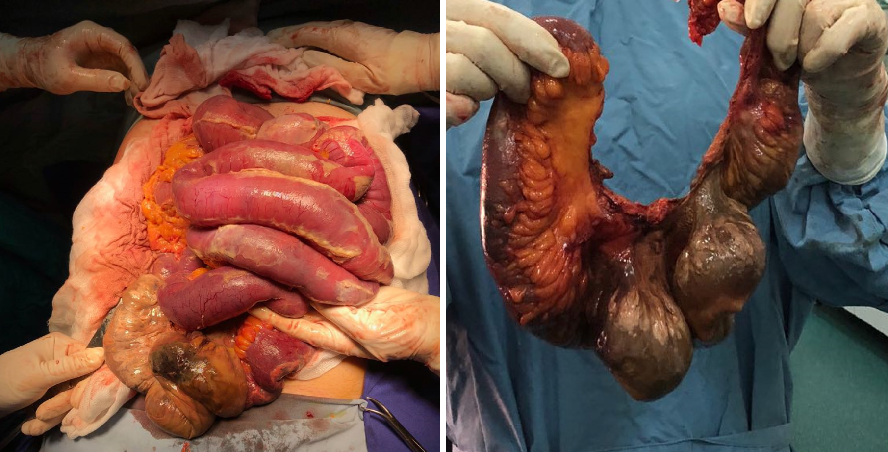
Figure 5 Ileum necrosis.
Macroscopic appearance of the resection piece.
- Citation: Șalaru DL, Adam CA, Marcu DTM, Șimon IV, Macovei L, Ambrosie L, Chirita E, Sascau RA, Statescu C. Acute myocardial infarction and extensive systemic thrombosis in thrombotic thrombocytopenic purpura: A case report and review of literature. World J Clin Cases 2021; 9(27): 8104-8113
- URL: https://www.wjgnet.com/2307-8960/full/v9/i27/8104.htm
- DOI: https://dx.doi.org/10.12998/wjcc.v9.i27.8104


