©The Author(s) 2021.
World J Clin Cases. Sep 26, 2021; 9(27): 8071-8081
Published online Sep 26, 2021. doi: 10.12998/wjcc.v9.i27.8071
Published online Sep 26, 2021. doi: 10.12998/wjcc.v9.i27.8071
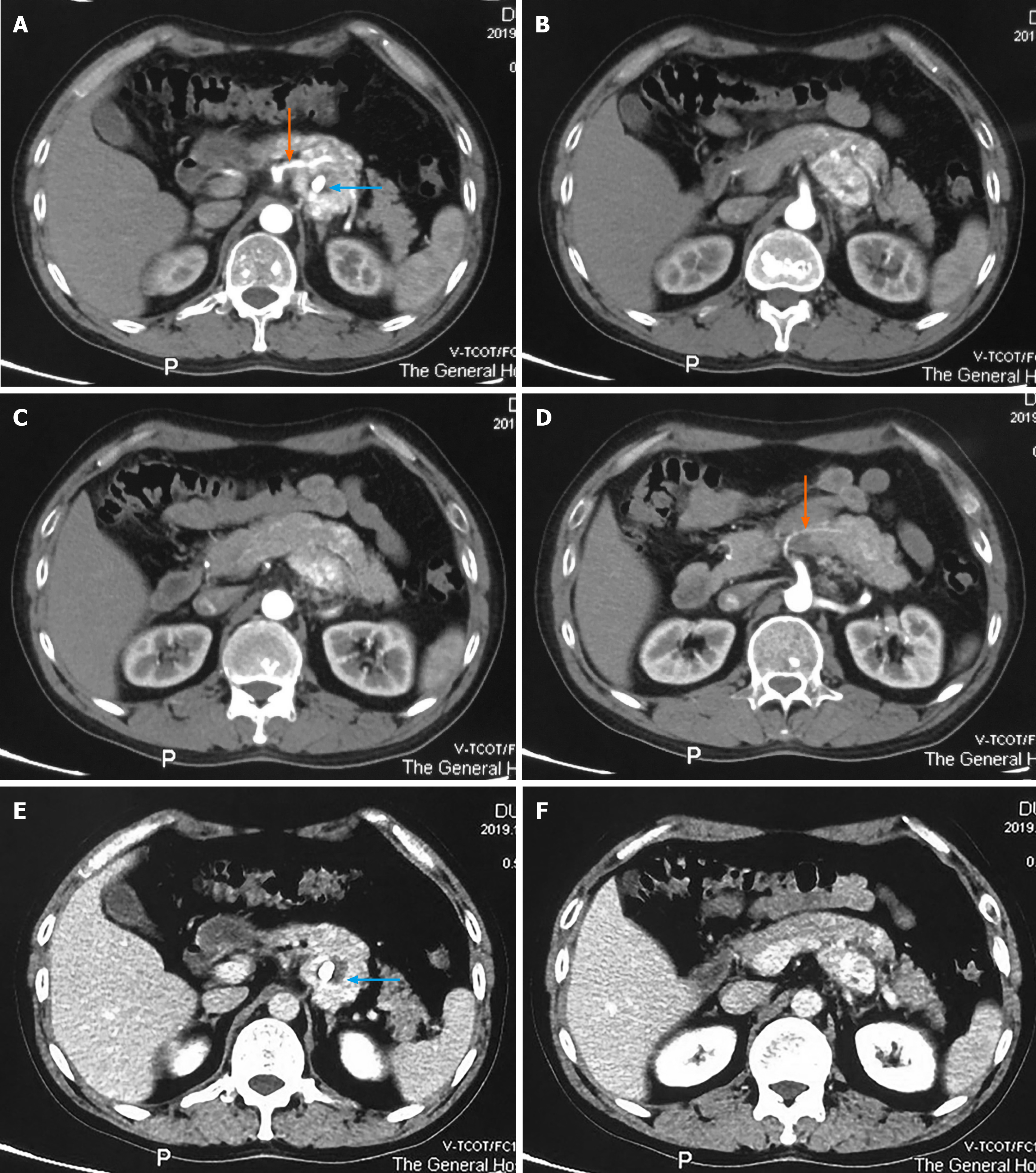
Figure 1 Abdominal contrast enhanced computed tomography findings.
A-D: The tumor shows marked enhancement during the arterial phase, the orange arrows show the vessels supplying the tumor from the superior mesenteric artery, and nodular calcification is observed inside the tumor (blue arrow); E and F: Enhancement is maintained, to a certain degree, during the venous phase, and nodular calcification was observed inside the tumor (blue arrow).
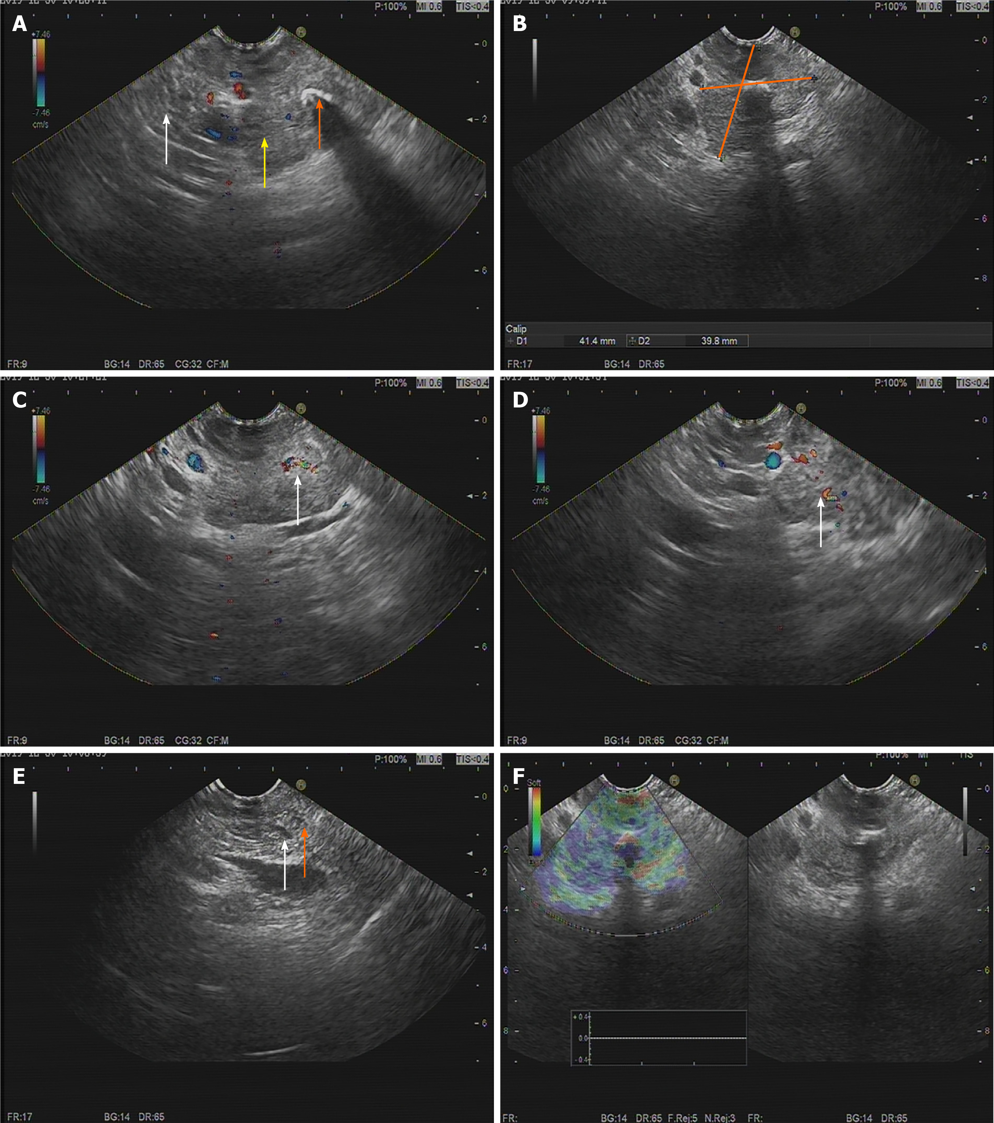
Figure 2 Endoscopic ultrasonography findings.
A: The tumor (yellow arrow) has an obscure boundary with the pancreas (white arrow), and nodular calcification can be seen inside the tumor (orange arrow); B: The tumor is 41.4 mm × 39.8 mm in size; C and D: A modest blood flow signal was detected inside the tumor (white arrow); E: No dilation of the bile duct (white arrow) and pancreatic duct (orange arrow); F: Elastography revealed the soft texture of the tumor.
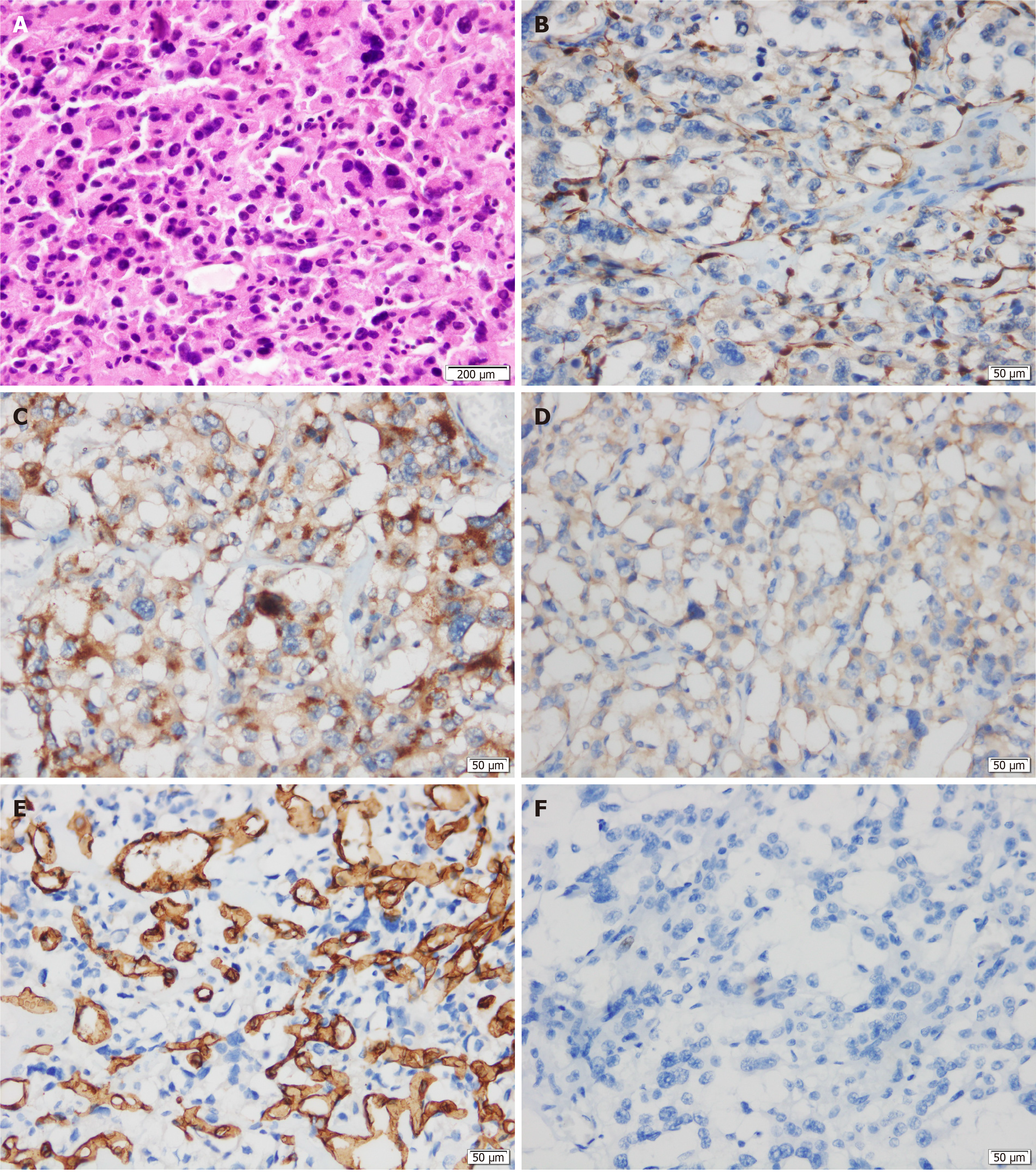
Figure 3 Histological and immunohistochemical examinations of the resected tumor.
A: Hematoxylin-eosin staining shows the classic Zellballen pattern; B: The sustentacular cells of tumor tissue are positive for S-100 protein; C: The neoplastic cells are positive for chromogranin A; D: The neoplastic cells are positive for synaptophysin; E: Positive staining for CD34 in the surrounding fibrous septa highlights abundant vascularization inside the septa; F: Ki-67 assessment (+, 2%).
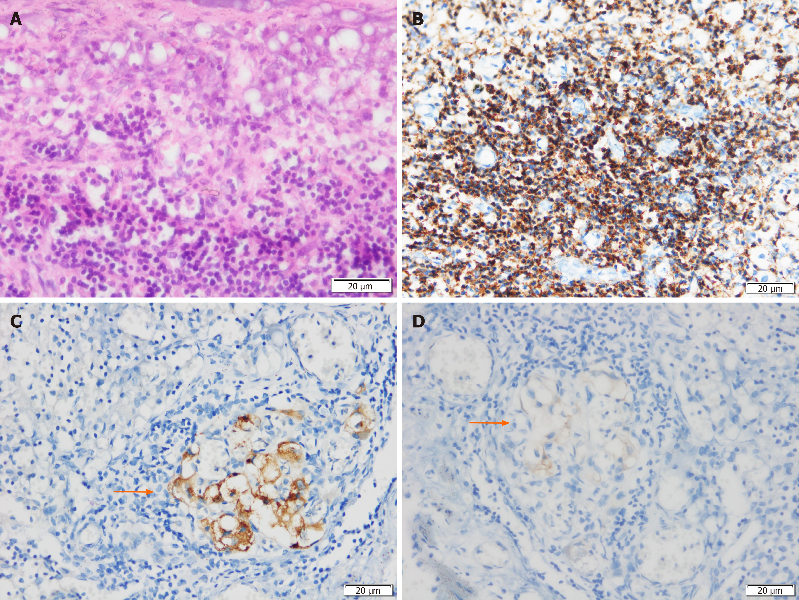
Figure 4 Histological and immunohistochemical examinations of the resected pancreatic draining lymph nodes.
A: Lymphatic tissue observation by hematoxylin-eosin staining; B: Positive staining for leukocyte common antigen confirms lymphatic tissue; C: The resected lymph nodes are positive for chromogranin A; D: The resected lymph nodes are positive for synaptophysin.
- Citation: Jiang CN, Cheng X, Shan J, Yang M, Xiao YQ. Primary pancreatic paraganglioma harboring lymph node metastasis: A case report. World J Clin Cases 2021; 9(27): 8071-8081
- URL: https://www.wjgnet.com/2307-8960/full/v9/i27/8071.htm
- DOI: https://dx.doi.org/10.12998/wjcc.v9.i27.8071


