Copyright
©The Author(s) 2021.
World J Clin Cases. Sep 16, 2021; 9(26): 7901-7908
Published online Sep 16, 2021. doi: 10.12998/wjcc.v9.i26.7901
Published online Sep 16, 2021. doi: 10.12998/wjcc.v9.i26.7901
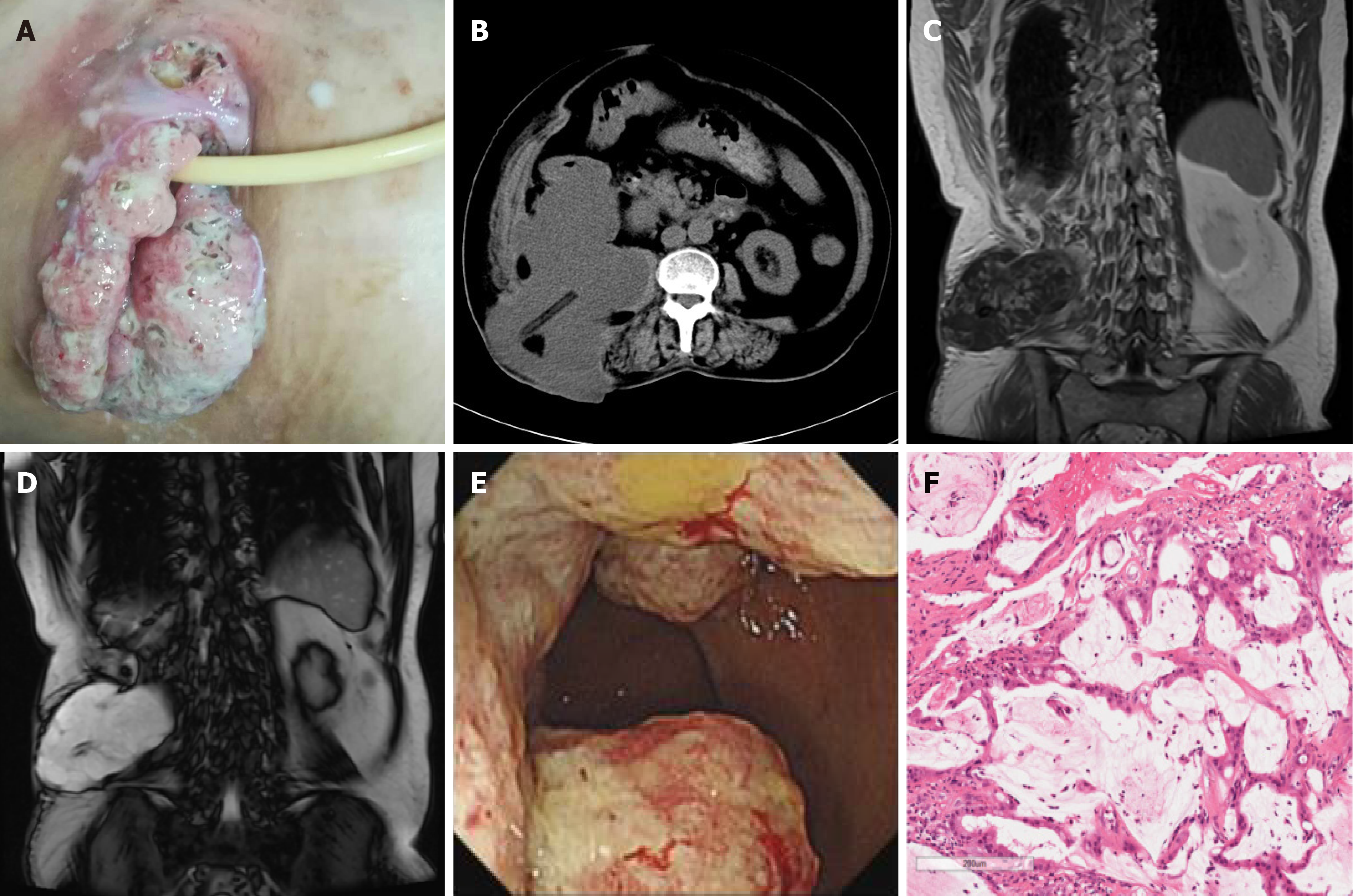
Figure 1 Case 1.
A: The incision of the right waist was cracked and inflamed; B: Abdominal computed tomography showed a mass between the psoas muscle and the ascending colon, and a drainage tube was visible; C and D: Abdominal MRI showed an irregular mass with a long T1 and long T2 signal in the right posterior abdominal para-vertebral region; E: A horseshoe-shaped mass in the ascending colon; F: Pathology of the colonic mucinous adenocarcinoma (hematoxylin-eosin staining, 20 ×).
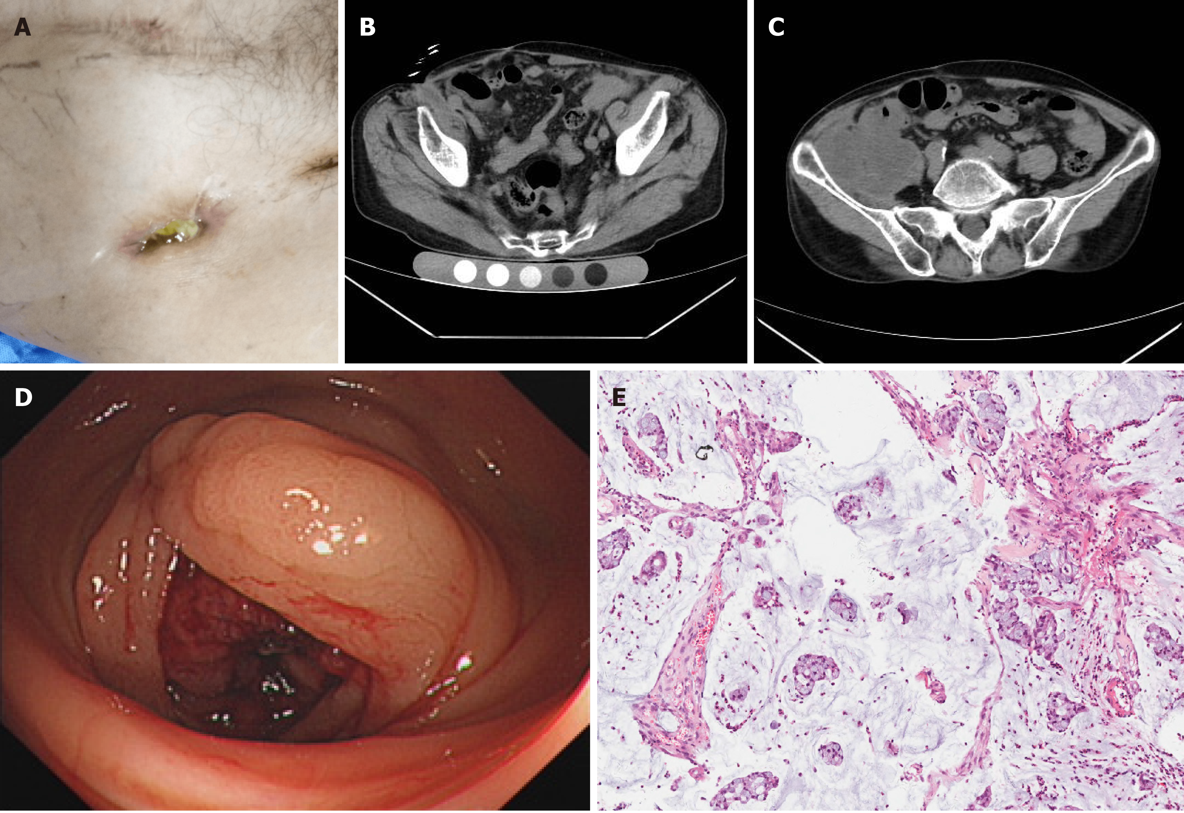
Figure 2 Case 2.
A: A sinus approximately 2 cm in size in the right inguinal region; B: The sinus on abdominal computed tomography; C: A mass in the right iliac fossa with unclear local and cecal boundaries; D: A horseshoe-shaped mass in the ascending colon; E: Pathology of the colon mucinous adenocarcinoma (hematoxylin-eosin staining, 20 ×).
Figure 3 Case 3.
A: Abdominal computed tomography scan showed a mass that extended to the subcutaneous tissue, and the boundary with the intestine was unclear; B: A mass in the ascending colon in the lumen; C: Pathological examination confirmed colon mucinous adenocarcinoma (hematoxylin-eosin staining, 20 ×).
- Citation: Han SZ, Wang R, Wen KM. Delayed diagnosis of ascending colon mucinous adenocarcinoma with local abscess as primary manifestation: Report of three cases. World J Clin Cases 2021; 9(26): 7901-7908
- URL: https://www.wjgnet.com/2307-8960/full/v9/i26/7901.htm
- DOI: https://dx.doi.org/10.12998/wjcc.v9.i26.7901


