Copyright
©The Author(s) 2021.
World J Clin Cases. Sep 16, 2021; 9(26): 7818-7824
Published online Sep 16, 2021. doi: 10.12998/wjcc.v9.i26.7818
Published online Sep 16, 2021. doi: 10.12998/wjcc.v9.i26.7818
Figure 1 Bone marrow morphology.
A: Myeloproliferative actively and absence of erythroid hyperplasia; B: Large granular lymphocyte (arrowheads). A and B: Wright staining, 1000 ×.
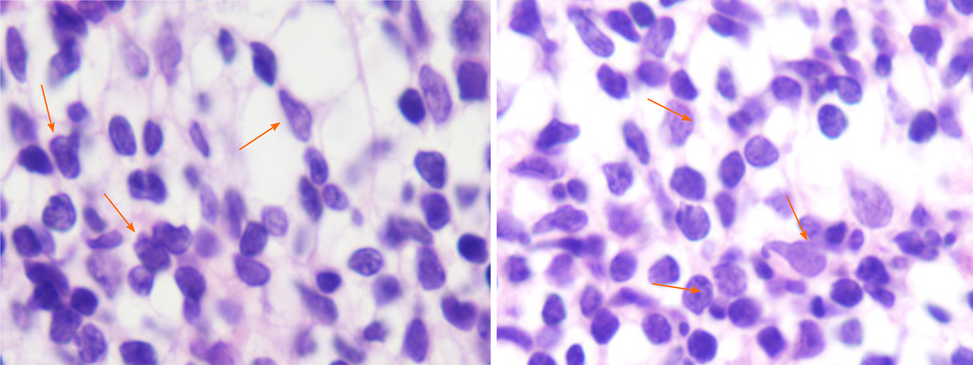
Figure 2 Histopathology of bone marrow tissue.
Large granular lymphocyte (arrowheads) (hematoxylin and eosin staining, 1000 ×).
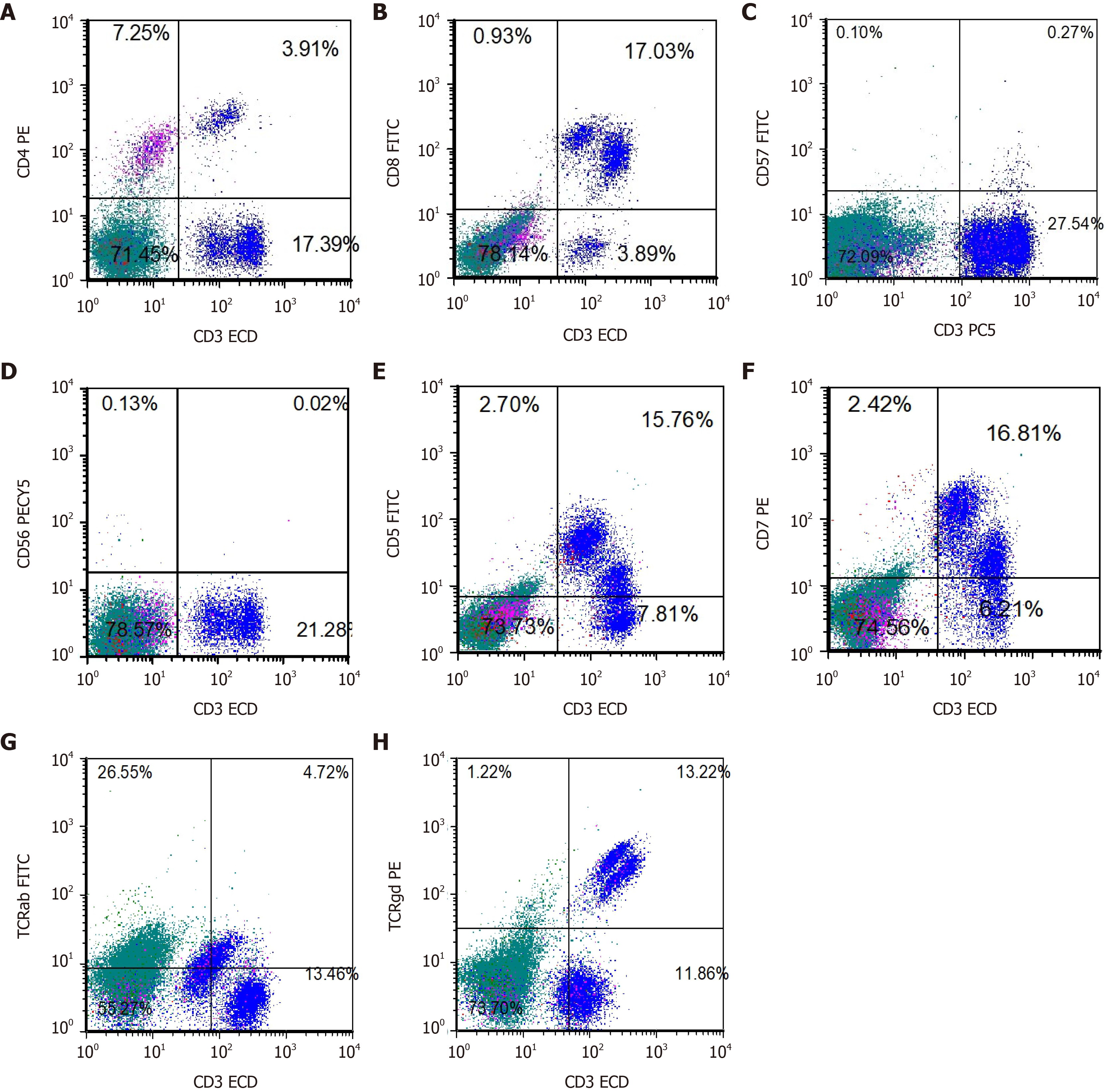
Figure 3 Immunophenotype of bone marrow by flow cytometry.
A: CD4 PE; B: CD8 fluorescein isothiocyanate (FITC); C: CD57 FITC; D: CD56 PECY5; E: CD5 FITC; F: CD7 PE; G: T-cell receptors ab FITC; H: TCRgd PE. TCR: T-cell receptors.
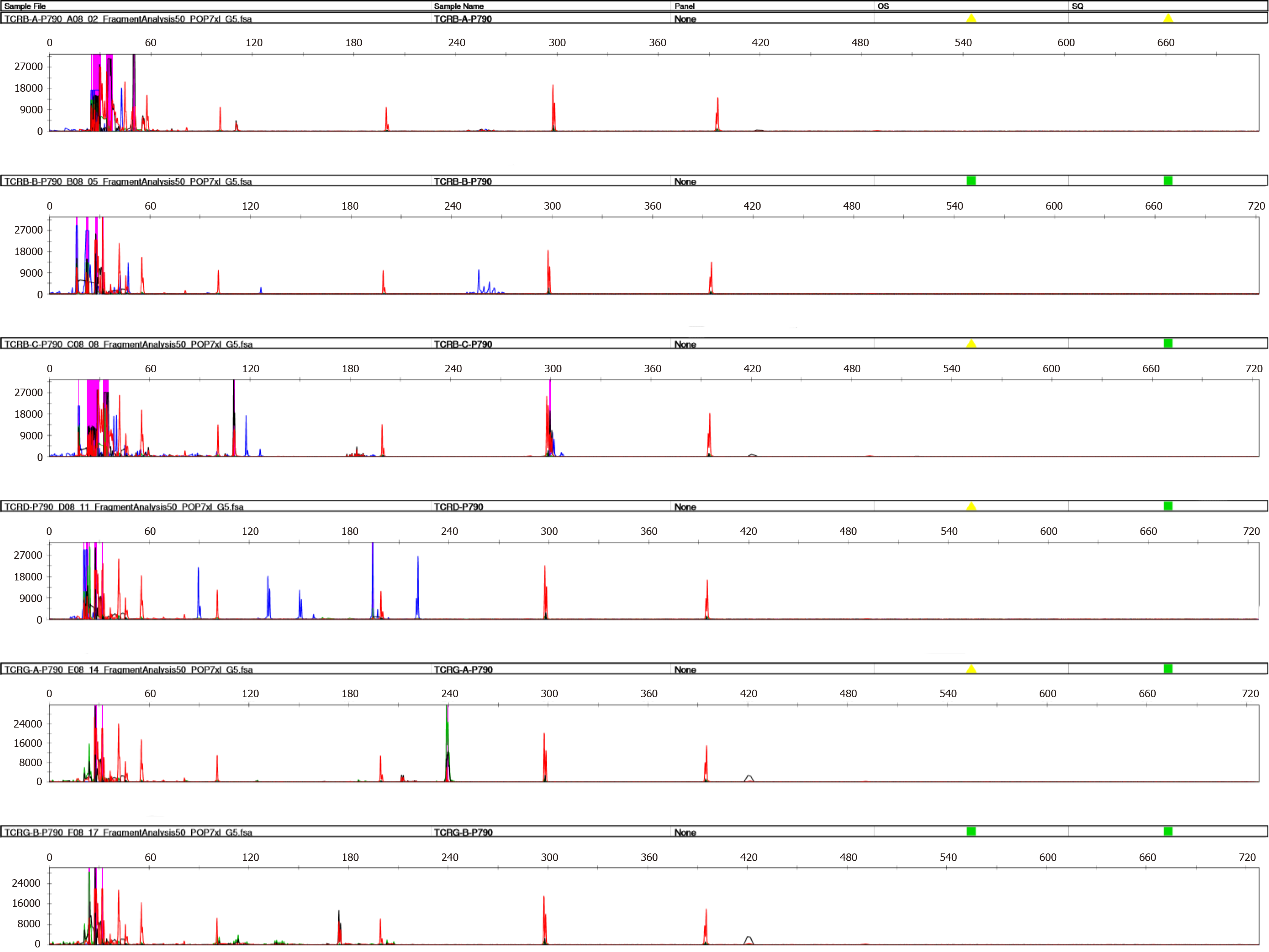
Figure 4 Monoclonal T-cell receptors rearrangement of bone marrow.
- Citation: Xiao PP, Chen XY, Dong ZG, Huang JM, Wang QQ, Chen YQ, Zhang Y. Treatment for CD57-negative γδ T-cell large granular lymphocytic leukemia with pure red cell aplasia: A case report. World J Clin Cases 2021; 9(26): 7818-7824
- URL: https://www.wjgnet.com/2307-8960/full/v9/i26/7818.htm
- DOI: https://dx.doi.org/10.12998/wjcc.v9.i26.7818


