©The Author(s) 2021.
World J Clin Cases. Aug 16, 2021; 9(23): 6639-6653
Published online Aug 16, 2021. doi: 10.12998/wjcc.v9.i23.6639
Published online Aug 16, 2021. doi: 10.12998/wjcc.v9.i23.6639
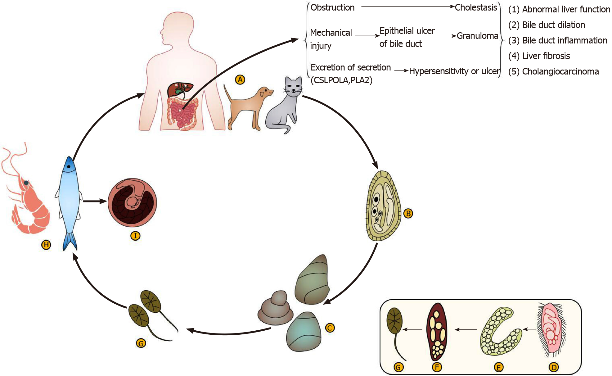
Figure 1 The life history of Clonorchis sinensis.
A: Ultimate host; B: Egg; C: The first intermediate host; D: Miracidium; E: Sporocyst; F: Rediae; G: Cercaria; H: The second intermediate host; I: Cysticercus.
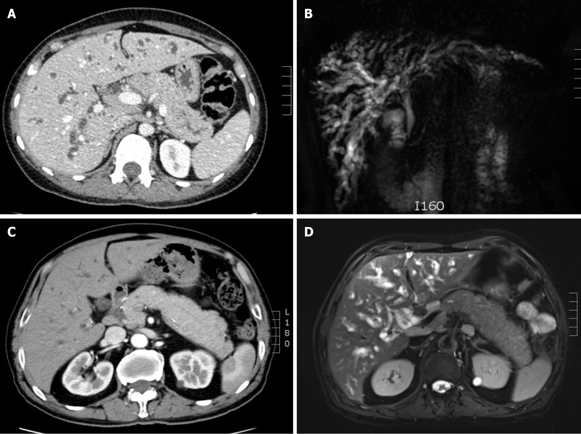
Figure 2 Images.
A: Computed tomography (CT) image of a woman. The intrahepatic bile duct was diffusely dilated, the pancreas was full, the pancreatic duct in the tail of the body was slightly dilated, and multiple enlarged lymph nodes in the hilar and retroperitoneum were visible; B: Magnetic resonance cholangiopancreatography (MRCP) image of the woman. Intrahepatic bile ducts were dilated and had an uneven thickness, and the pancreas was full; C: CT image of a man. The intrahepatic bile duct was diffusely dilated, and the pancreas was full; D: MRCP image of the man. The intrahepatic and extrahepatic bile ducts and common bile ducts were dilated, and enlarged peripancreatic lymph nodes were visible.
Figure 3 Pathological pictures.
A: Massive bile duct hyperplasia; B: Eosinophils and plasma cells; C: Immunoglobulin G 4.
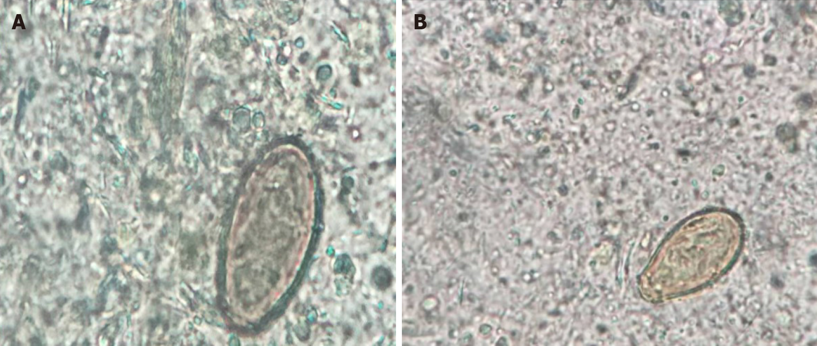
Figure 4 Clonorchis sinensis eggs in stool.
A: Woman; B: Man.
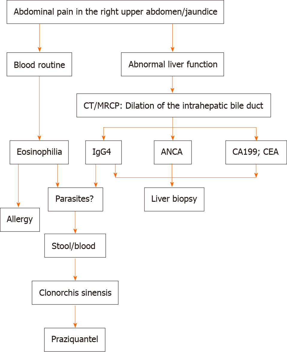
Figure 5 Flow diagram of diagnosis.
IgG4: Immunoglobulin G 4; ANCA: Anti-neutrophil cytoplasmic antibody; CEA: Carcinoembryonic antigen; CT: Computed tomography; MRCP: Magnetic resonance cholangiopancreatography.
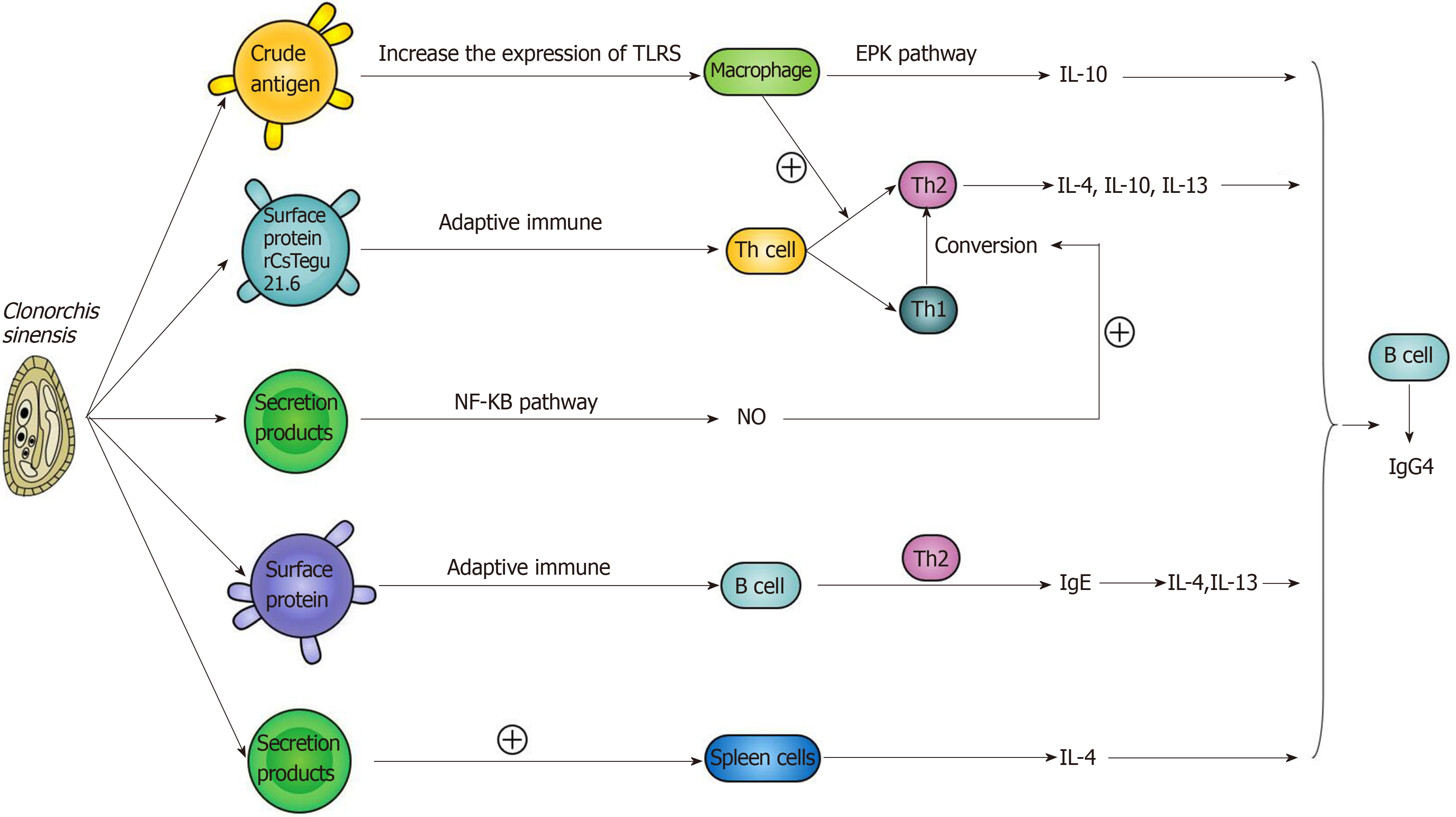
Figure 6 The related mechanisms between Clonorchis sinensis and immunoglobulin G4.
TLRS: Toll-like receptors; EPK: Eukaryotic protein kinase; IL: Interleukin; IgE: Immunoglobulins E; NF-kB: Nuclear factor kappa B.
- Citation: Zhang XH, Huang D, Li YL, Chang B. Novel mechanism of hepatobiliary system damage and immunoglobulin G4 elevation caused by Clonorchis sinensis infection. World J Clin Cases 2021; 9(23): 6639-6653
- URL: https://www.wjgnet.com/2307-8960/full/v9/i23/6639.htm
- DOI: https://dx.doi.org/10.12998/wjcc.v9.i23.6639


