©The Author(s) 2021.
World J Clin Cases. Aug 6, 2021; 9(22): 6418-6427
Published online Aug 6, 2021. doi: 10.12998/wjcc.v9.i22.6418
Published online Aug 6, 2021. doi: 10.12998/wjcc.v9.i22.6418
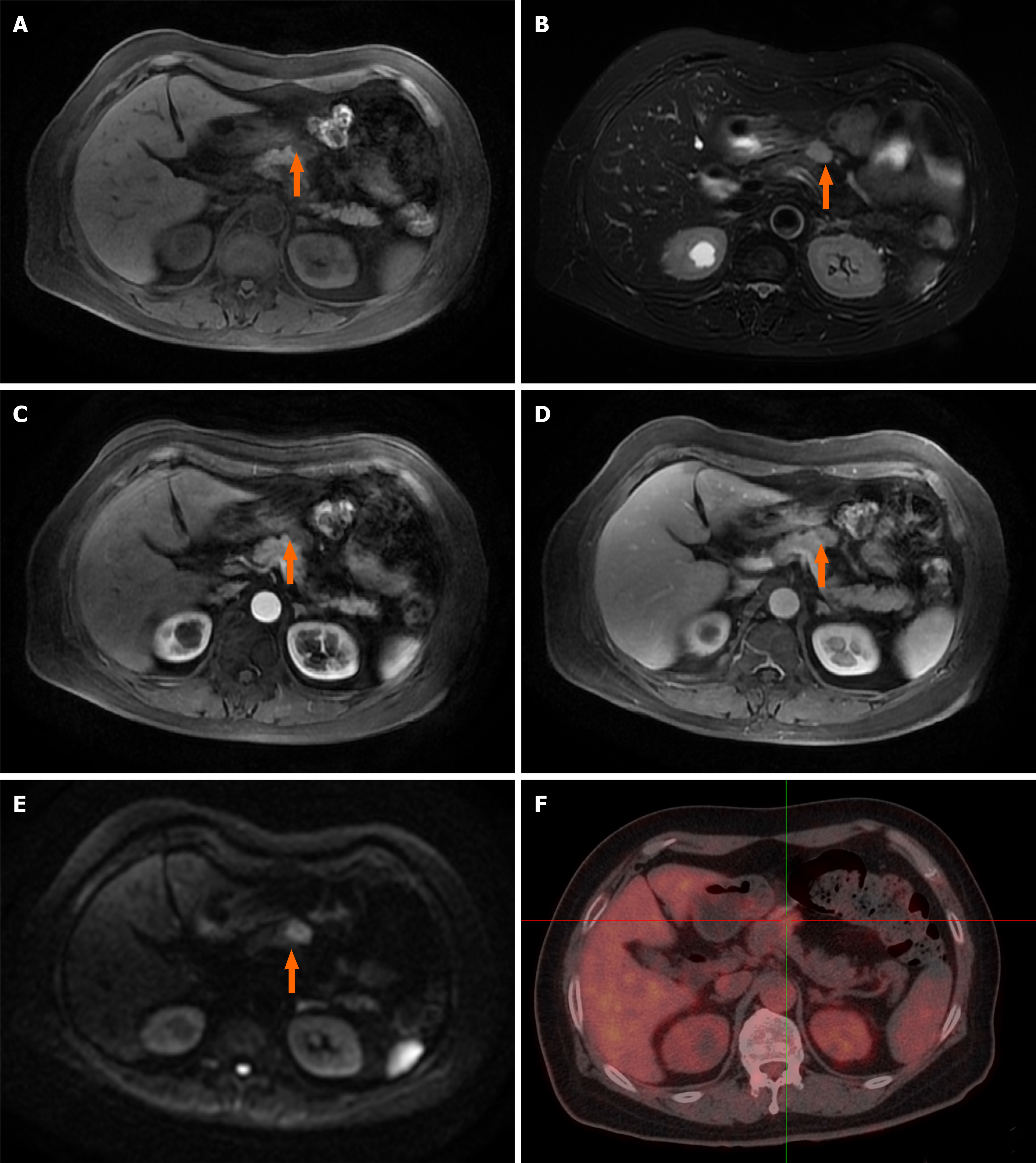
Figure 1 Dynamic contrast-enhanced magnetic resonance imaging and 18F-fluorodeoxyglucose positron emission tomography/computed tomography reveal one irregular lesion in the pancreatic neck.
A: The lesion of the pancreatic neck (orange arrow) was hyperintense on T1-weighted imaging; B: The irregular lesion (orange arrow) was slightly hyperintense on T2-weighted imaging; C: Arterial phase imaging revealed slight enhancement of the pancreatic lesion (orange arrow); D: The lesion (orange arrow) showed persistent enhancement in venous phase imaging; E: The pancreatic mass (orange arrow) had significant hyperintensity in diffusion-weighted imaging; F: 18F-fluorodeoxyglucose positron emission tomography/computed tomography revealed a hypermetabolic pancreatic neck nodule measuring 2.3 cm × 1.4 cm (SUVmax = 3.87).
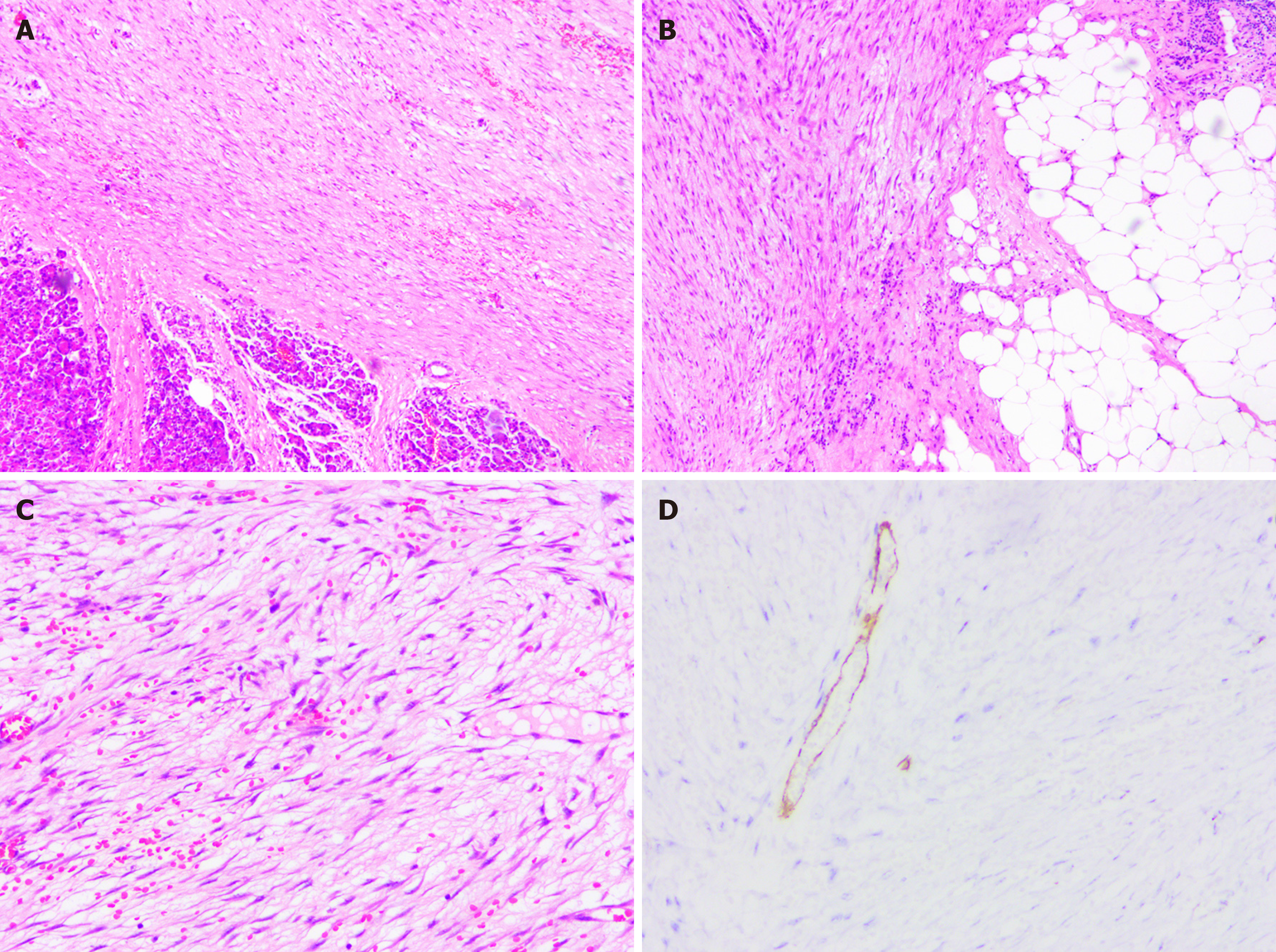
Figure 2 The pathological findings of the resected specimen revealed inflammatory myofibroblastic tumor.
A-C: Histological images showed mixed components of dense myofibroblastic tissues and few inflammatory cells, with neoplastic cells infiltrating the surrounding fat tissue (hematoxylin and eosin staining); D: Immunohistochemical studies showed positivity for smooth muscle actin.
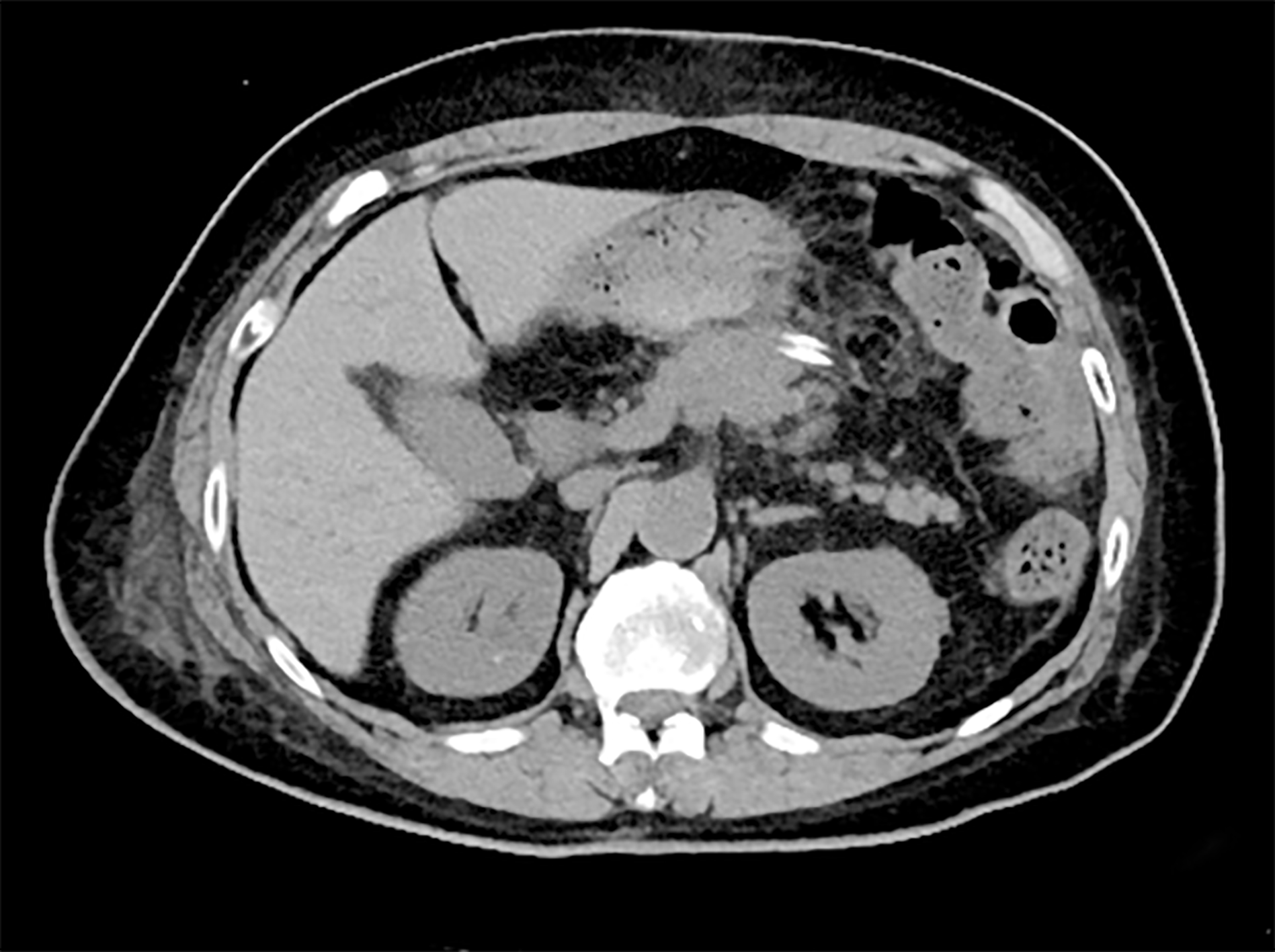
Figure 3 Ten days after surgical resection, computed tomography showed that the pancreatic neck inflammatory myofibroblastic tumor was enucleated, and the tissue of the pancreas remained intact.
- Citation: Chen ZT, Lin YX, Li MX, Zhang T, Wan DL, Lin SZ. Inflammatory myofibroblastic tumor of the pancreatic neck: A case report and review of literature. World J Clin Cases 2021; 9(22): 6418-6427
- URL: https://www.wjgnet.com/2307-8960/full/v9/i22/6418.htm
- DOI: https://dx.doi.org/10.12998/wjcc.v9.i22.6418


