Copyright
©The Author(s) 2021.
World J Clin Cases. Jul 26, 2021; 9(21): 6026-6031
Published online Jul 26, 2021. doi: 10.12998/wjcc.v9.i21.6026
Published online Jul 26, 2021. doi: 10.12998/wjcc.v9.i21.6026
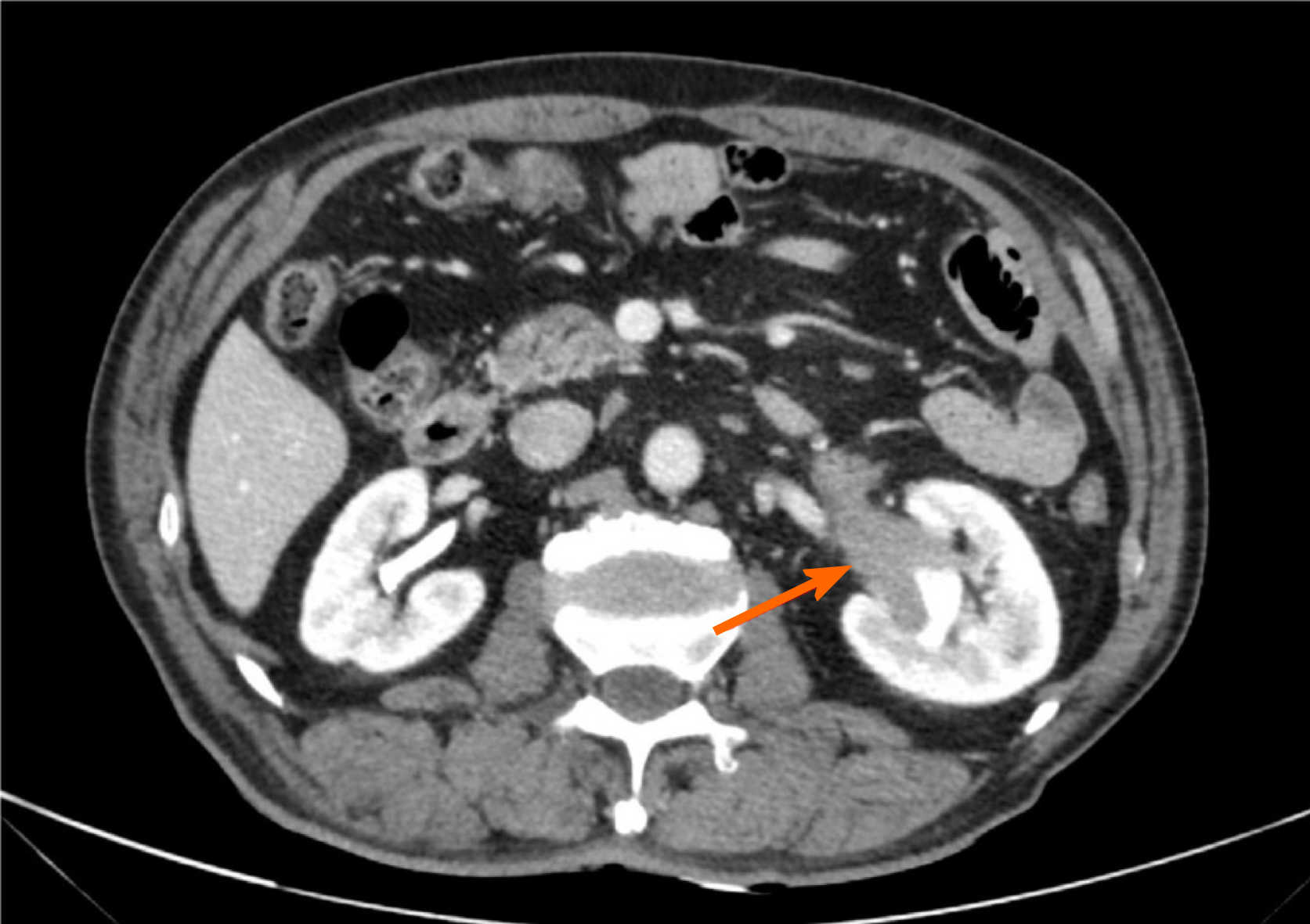
Figure 1 Abdominal enhanced computed tomography scan showed soft tissue masses in the left renal pelvis and the beginning of the left ureter, with uniform density and moderate enhancement.
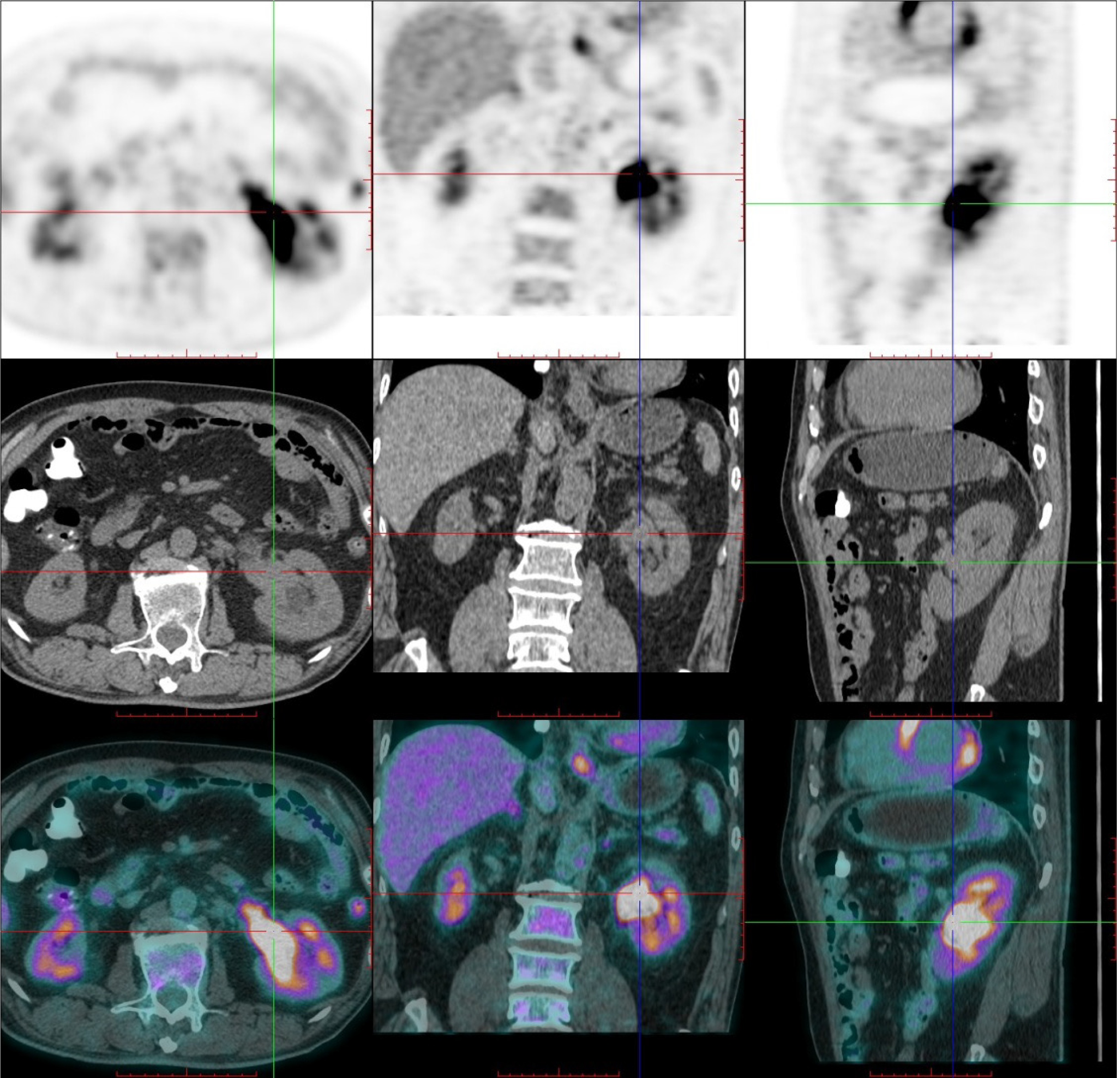
Figure 2 Delayed positron emission tomography/computed tomography imaging 1.
5 h later revealed a high uptake of fluorodeoxyglucose in the renal pelvic tumor.
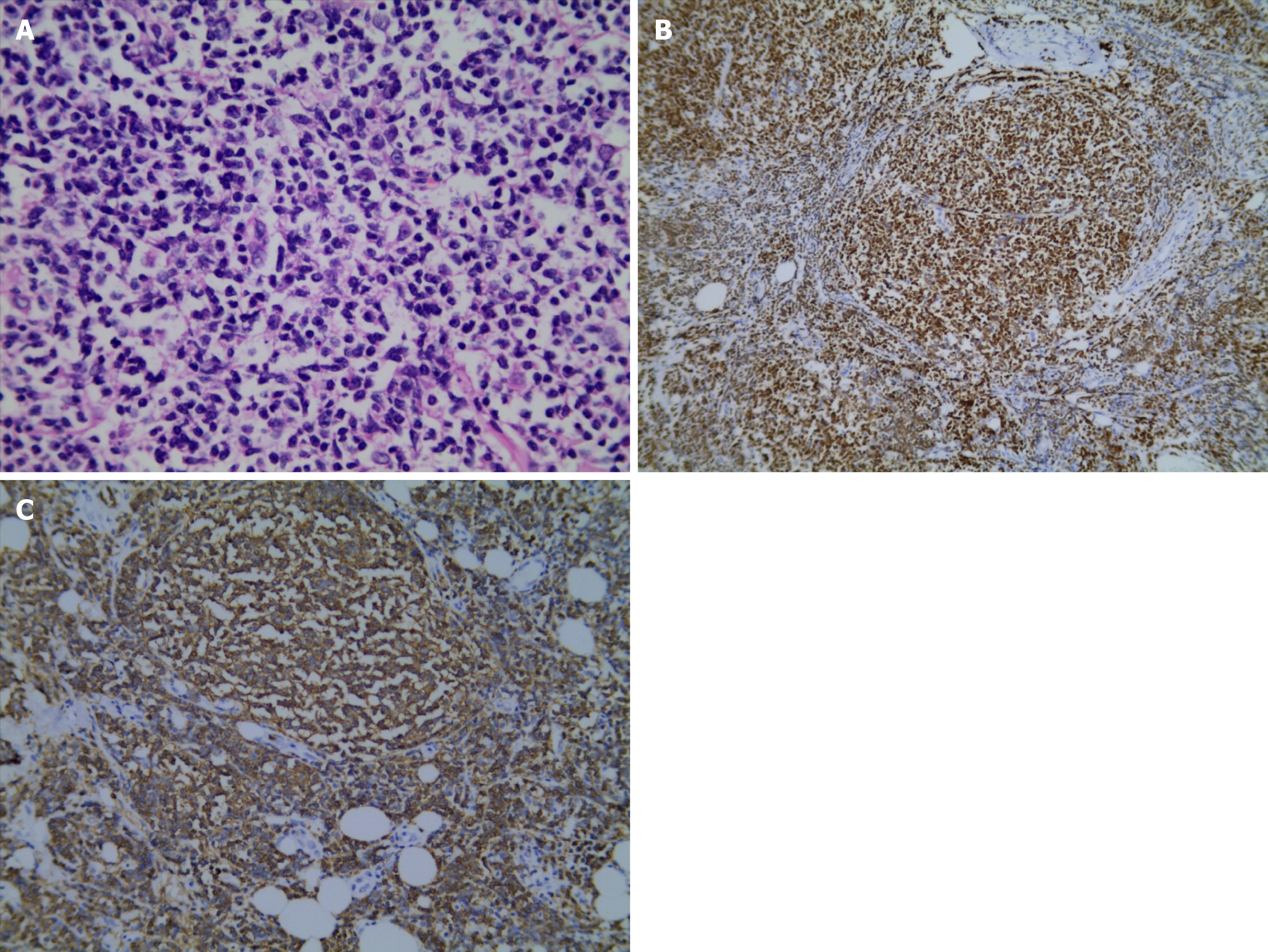
Figure 3 Pathology of the renal pelvis.
A: Involvement of ureter by follicular lymphoma (hematoxylin and eosin stain × 400); B: Immunohistochemical staining with Bcl-2 (× 100); C: Immunohistochemical staining with CD20 (× 200).
- Citation: Shen XZ, Lin C, Liu F. Primary follicular lymphoma in the renal pelvis: A rare case report. World J Clin Cases 2021; 9(21): 6026-6031
- URL: https://www.wjgnet.com/2307-8960/full/v9/i21/6026.htm
- DOI: https://dx.doi.org/10.12998/wjcc.v9.i21.6026


