Copyright
©The Author(s) 2021.
World J Clin Cases. Jul 6, 2021; 9(19): 5252-5258
Published online Jul 6, 2021. doi: 10.12998/wjcc.v9.i19.5252
Published online Jul 6, 2021. doi: 10.12998/wjcc.v9.i19.5252
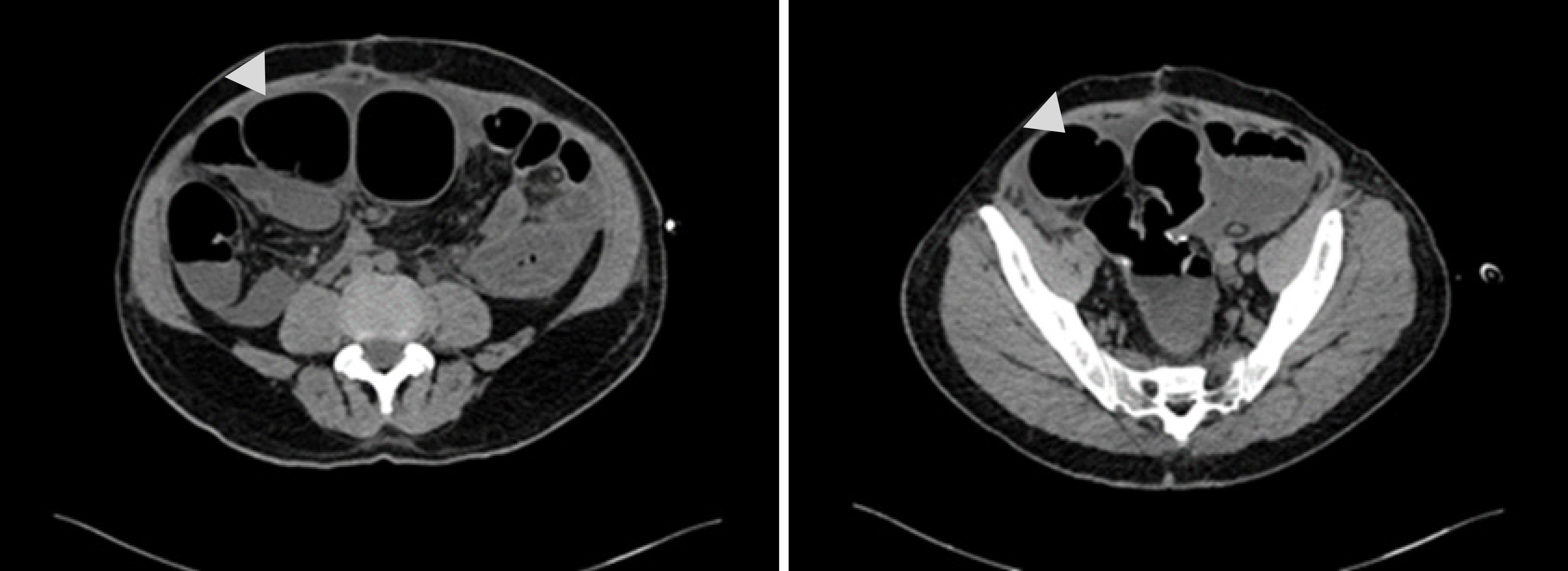
Figure 1 Abdominal computed tomography revealed sever intestinal distension of the indwelling colon.
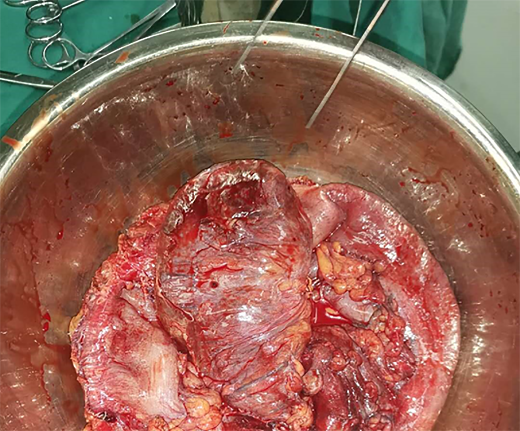
Figure 2 Congested and swollen colon were found during the operation.
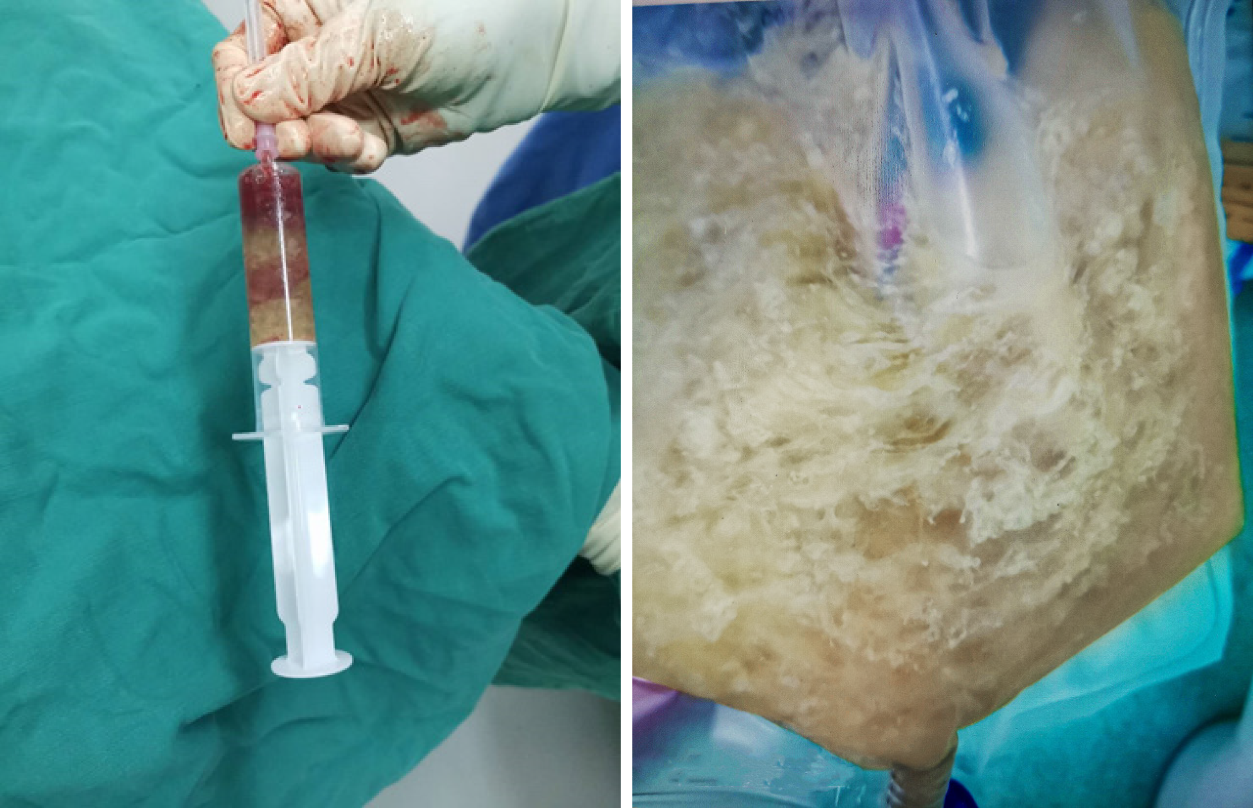
Figure 3 Large amounts of viscous liquid drained from the indwelling colon.
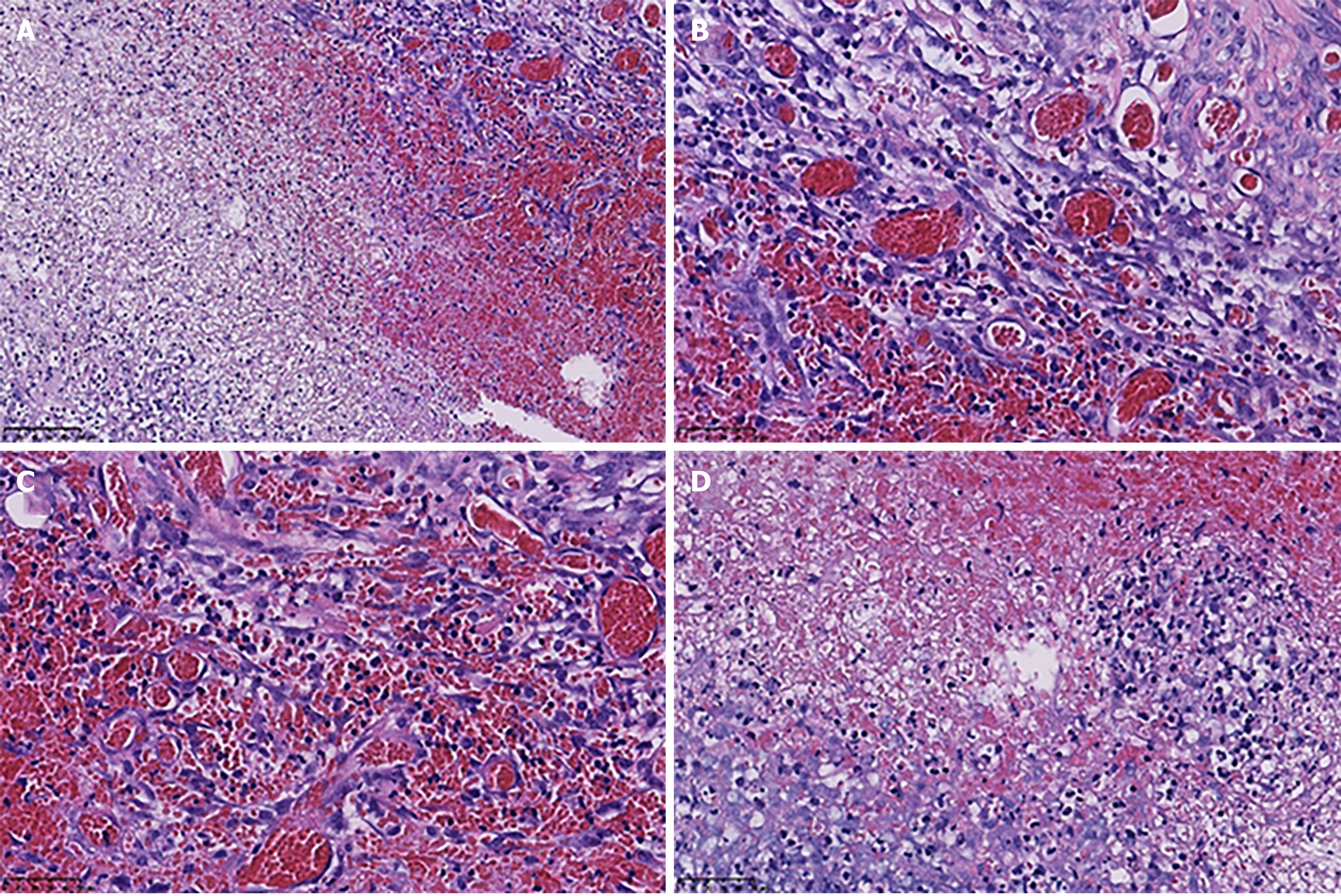
Figure 4 Pathological images of the distension colon.
Colonic wall congestion and necrosis with hyperplasia of granulation tissue. A: Hematoxylin and eosin (HE) staining, × 200; B: HE staining, × 400; C: HE staining, × 400; D: HE staining, × 400.
- Citation: Zhuang ZX, Wei MT, Yang XY, Zhang Y, Zhuang W, Wang ZQ. Long-term outcome of indwelling colon observed seven years after radical resection for rectosigmoid cancer: A case report. World J Clin Cases 2021; 9(19): 5252-5258
- URL: https://www.wjgnet.com/2307-8960/full/v9/i19/5252.htm
- DOI: https://dx.doi.org/10.12998/wjcc.v9.i19.5252


