©The Author(s) 2021.
World J Clin Cases. Jun 26, 2021; 9(18): 4627-4636
Published online Jun 26, 2021. doi: 10.12998/wjcc.v9.i18.4627
Published online Jun 26, 2021. doi: 10.12998/wjcc.v9.i18.4627
Figure 1 Comparison of amygdala magnetic resonance imaging parameters in two groups.
The left and right amygdala volumes of Alzheimer’s disease patients were significantly lower than controls. A: Left amygdala volume; B: Right amygdala volume. AD: Alzheimer’s disease.
Figure 2 Amygdala volume in different dementia severity.
There was no significant difference in the left and right amygdala volume among the three subgroups (P > 0.05). A: Left amygdala volume; B: Right amygdale volume. AD: Alzheimer’s disease.
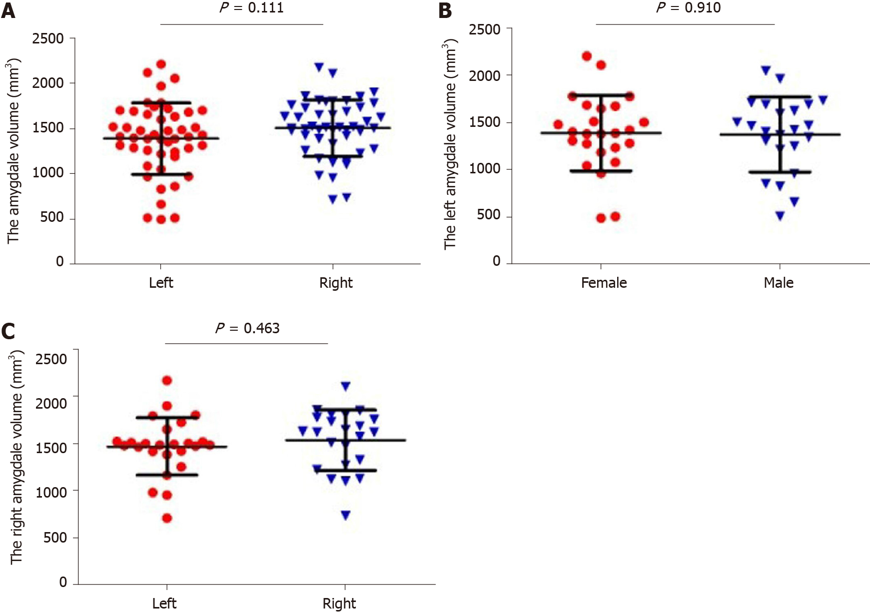
Figure 3 Amygdala volume between different sides and genders.
A: There was no significant difference between left and right amygdala (P > 0.05); B, C: There was no significant difference in amygdala volume between different genders (P > 0.05).
Figure 4 Amygdala volumes between Alzheimer's disease patients with and without mental symptoms.
The left and right amygdala volumes of Alzheimer's disease patients with mental symptoms were significantly smaller than patients without mental symptoms. A: Left amygdala volume; B: Right amygdala volume.
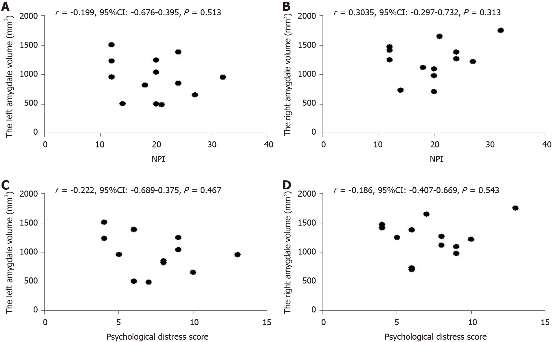
Figure 5 Correlation analyses between neuropsychiatric inventory score and bilateral amygdala.
A, B: There was no significant correlation between neuropsychiatric inventory scores and amygdala volumes (P > 0.05); C, D: There was no correlation between psychological distress scores of caregivers and amygdala volumes (P > 0.05).
Figure 6 Correlation between amygdala and hippocampus volume.
A: There was significant correlation between left amygdala and hippocampal volumes (P < 0.001); B: There was a significant correlation between the right amygdala volume and hippocampal volume (P < 0.001).
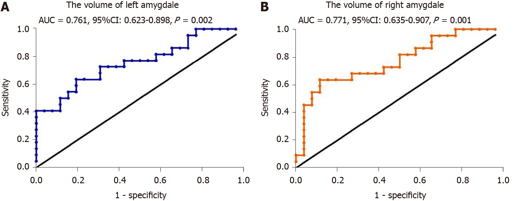
Figure 7 Receiver operating characteristic curve analysis of amygdala volume for diagnosis of Alzheimer’s disease.
A: Volume of left amygdala; B: Volume of right amygdala. AUC: Area under the curve.
- Citation: Wang DW, Ding SL, Bian XL, Zhou SY, Yang H, Wang P. Diagnostic value of amygdala volume on structural magnetic resonance imaging in Alzheimer’s disease. World J Clin Cases 2021; 9(18): 4627-4636
- URL: https://www.wjgnet.com/2307-8960/full/v9/i18/4627.htm
- DOI: https://dx.doi.org/10.12998/wjcc.v9.i18.4627


