©The Author(s) 2021.
World J Clin Cases. Apr 16, 2021; 9(11): 2627-2633
Published online Apr 16, 2021. doi: 10.12998/wjcc.v9.i11.2627
Published online Apr 16, 2021. doi: 10.12998/wjcc.v9.i11.2627
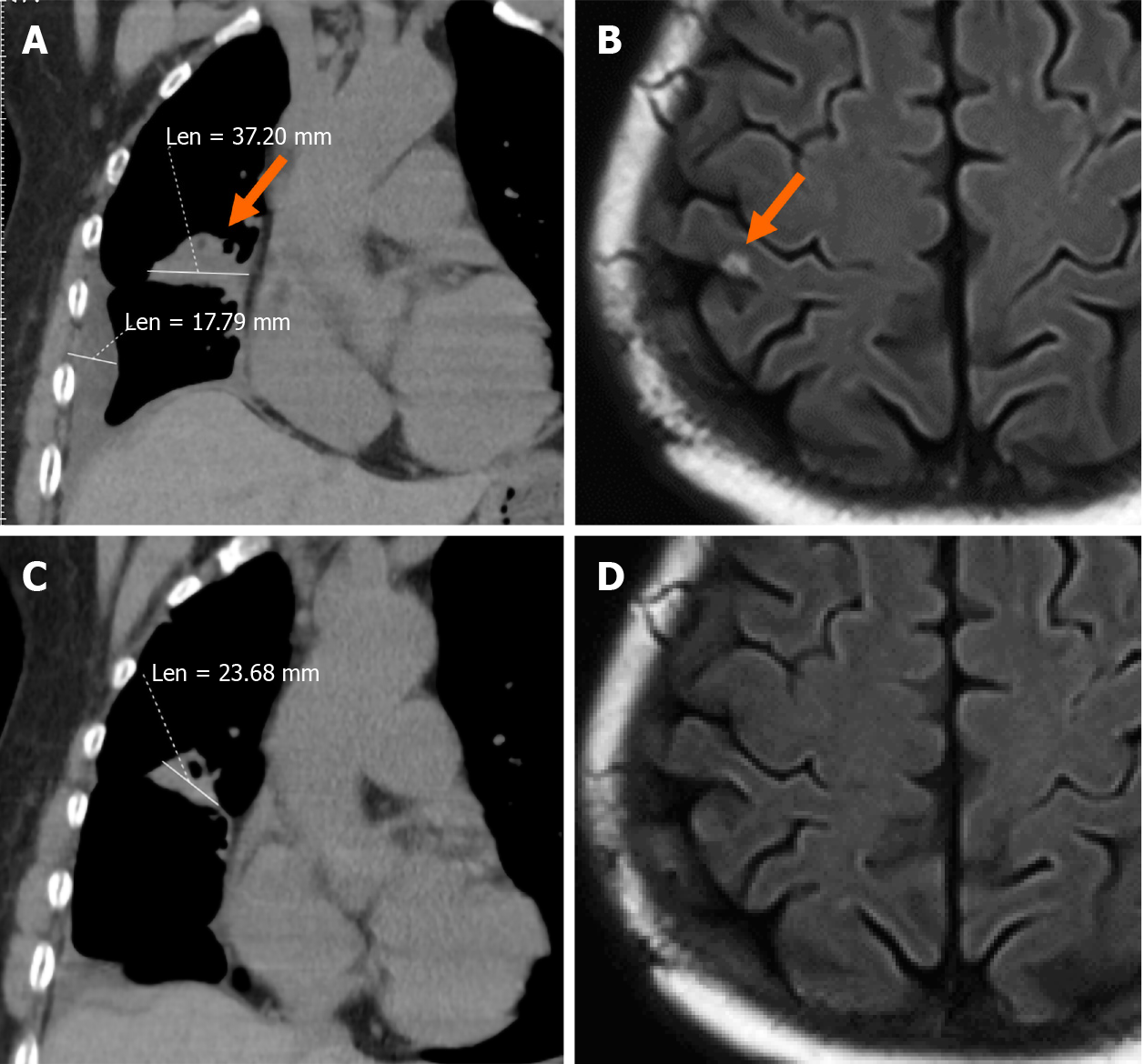
Figure 1 Chest computed tomography scan and brain magnetic resonance imaging from December 2017 to May 2018.
A: Chest computed tomography (CT) scan as baseline before beginning therapy in December 2017; B: Brain magnetic resonance imaging showed a metastatic neoplasm in December 2017; C: Chest CT scan after 2 mo of therapy in March 2018; D: The neoplasm had disappeared in May 2018.
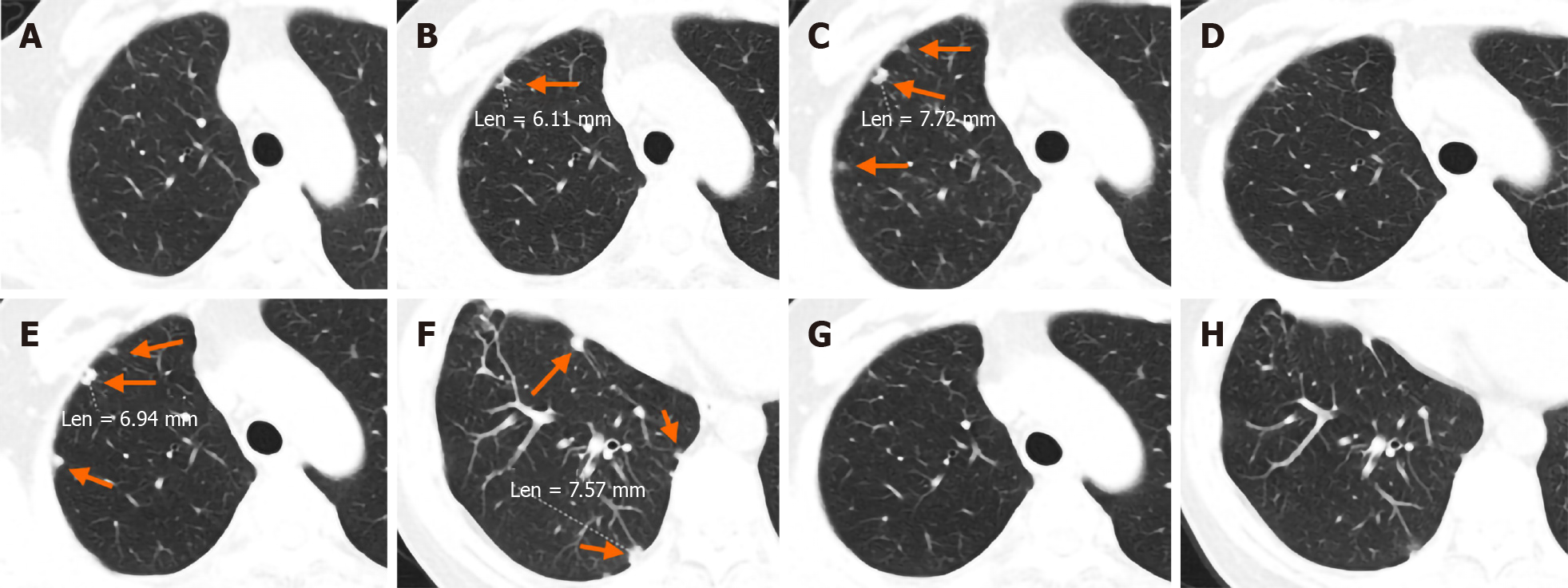
Figure 2 Chest computed tomography scans from October 2018 to November 2019.
A: Baseline image of the right pleura in October 2018; B: New foci in the pleura showed progressive disease in December 2018; C: The pleural metastatic lesions enlarged and increased in March 2019; D: The pleura foci almost disappeared after 2 mo of retreatment with osimertinib in May 2019; E and F: Secondary enlargement and increase of metastatic pleural foci in September and October 2019; G and H: The neoplasms disappeared again after 40 d tri-challenge with osimertinib in December 2019.
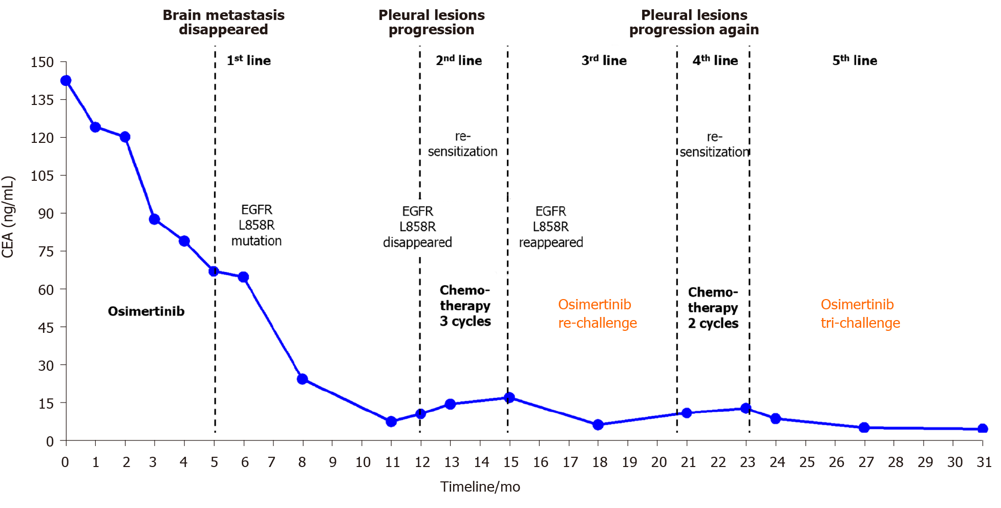
Figure 3 Entire treatment course with variations in carcinoembryonic antigen levels at each visit from the initial treatment to the present.
EGFR: Epidermal growth factor receptor; CEA: Carcinoembryonic antigen.
- Citation: Hu XY, Fei YC, Zhou WC, Zhu JM, Lv DL. Triple administration of osimertinib followed by chemotherapy for advanced lung adenocarcinoma: A case report. World J Clin Cases 2021; 9(11): 2627-2633
- URL: https://www.wjgnet.com/2307-8960/full/v9/i11/2627.htm
- DOI: https://dx.doi.org/10.12998/wjcc.v9.i11.2627


