©The Author(s) 2021.
World J Clin Cases. Apr 16, 2021; 9(11): 2576-2583
Published online Apr 16, 2021. doi: 10.12998/wjcc.v9.i11.2576
Published online Apr 16, 2021. doi: 10.12998/wjcc.v9.i11.2576
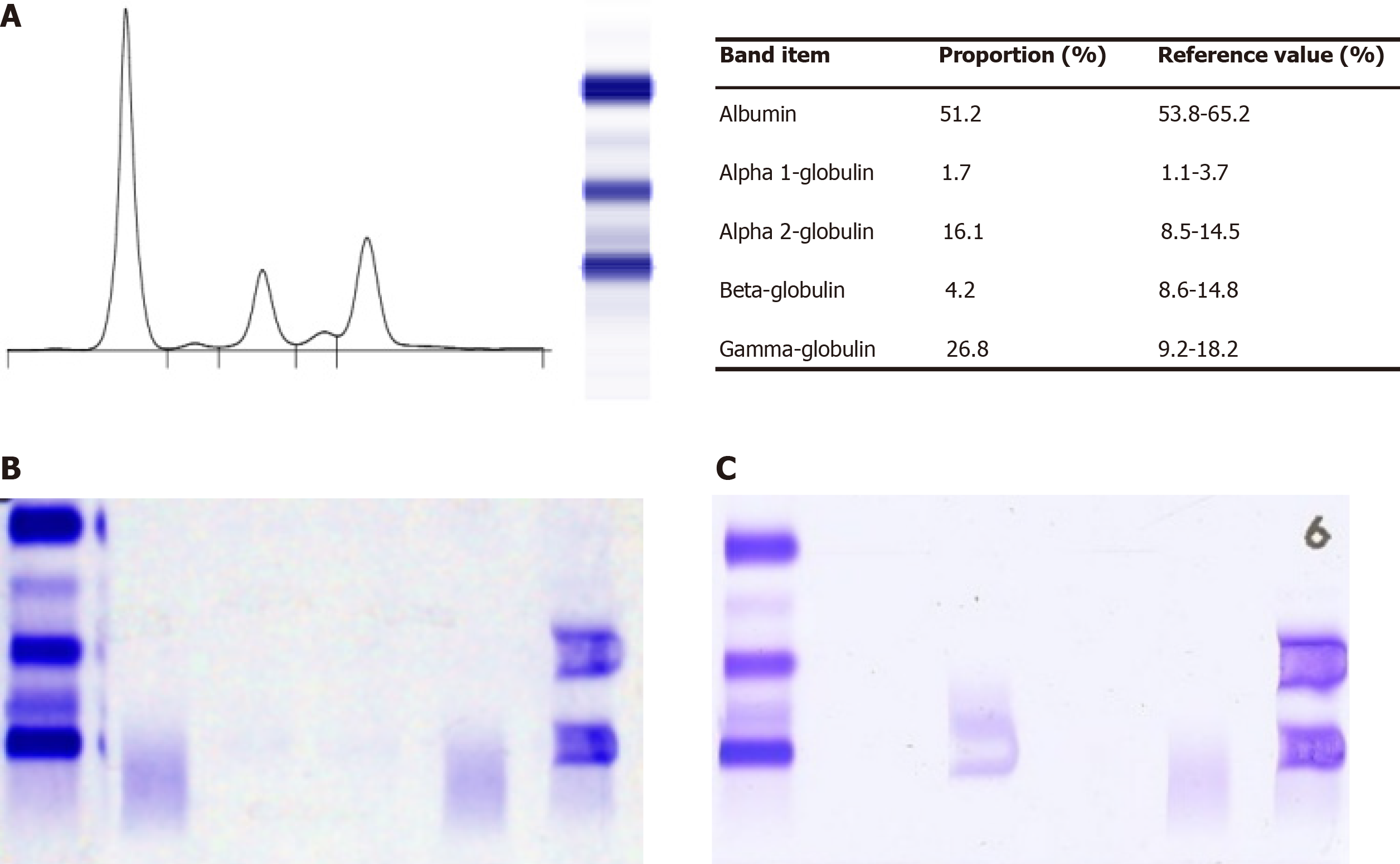
Figure 1 Immunoglobulin D-λ/λ myeloma proteins detected by electrophoresis (France, Sebia, HYDRASYS).
A: Serum protein electrophoresis showed two slight M-spikes that corresponded to alpha-2 and gamma globulins regions; B: Serum immuno-fixed electrophoresis [detected by anti-immunoglobulin (Ig) G, anti-IgA, anti-IgM, anti-κ, and anti-λ] presented two bands in anti-λ lane but no corresponding heavy chain band; C: Serum immuno-fixed electrophoresis (detected by anti-IgD, anti-IgE, anti-κ, and anti-λ) showed two M bands corresponding to IgD λ and free λ-light chains.
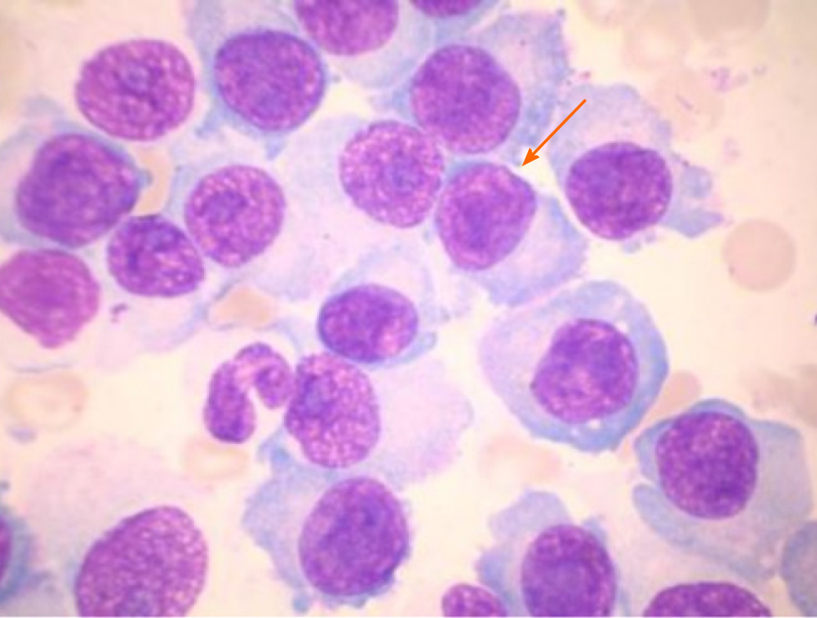
Figure 2 Bone marrow cytomorphologic examination (× 1000) showed a large number of immature plasma cells.
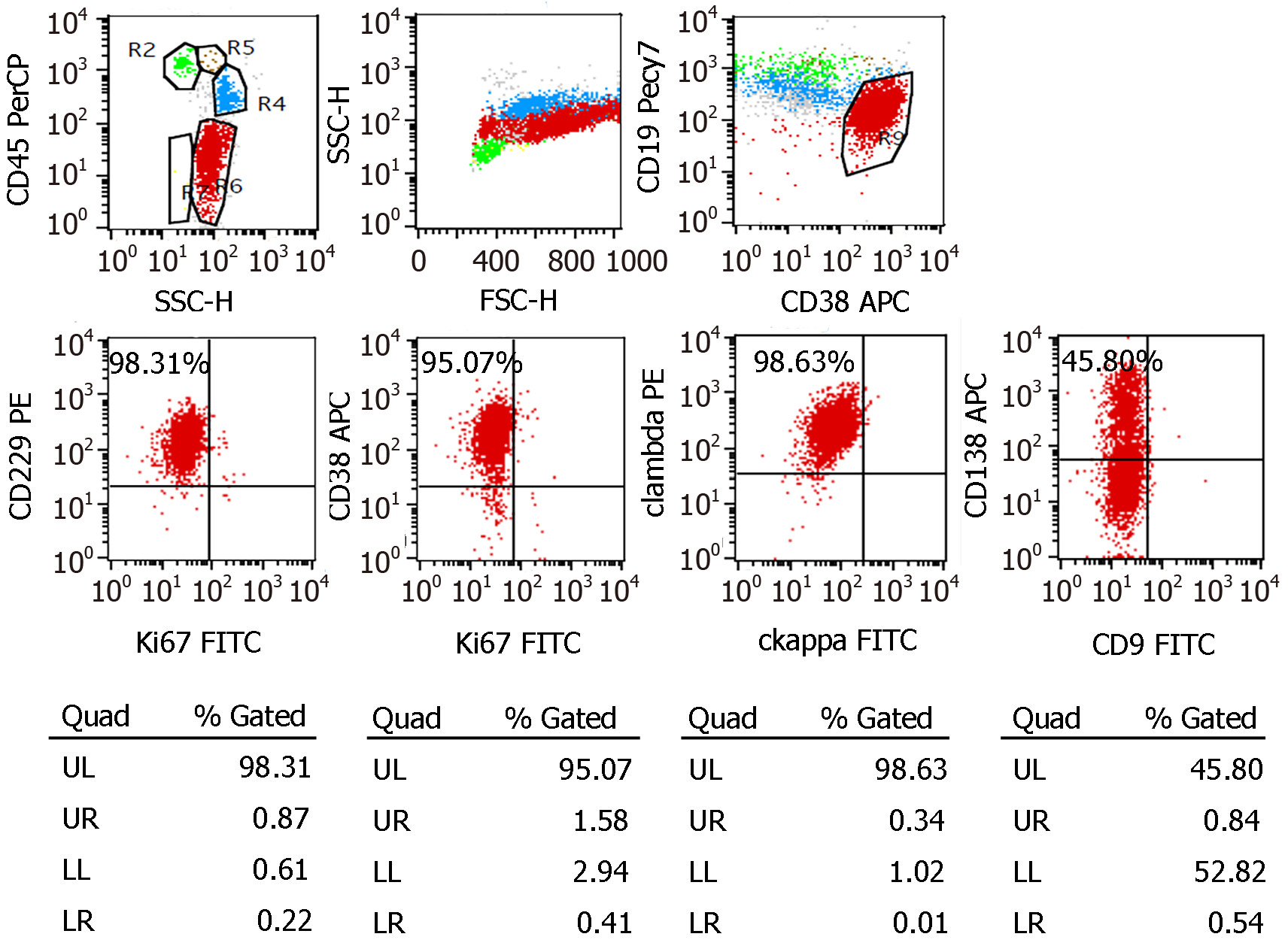
Figure 3 Flow cytometry (BD FACSDiva 8.
0.2) suggested an elevation of monoclonal plasma cells (70.12% of total nucleated red blood cells). All of the monoclonal plasma cells expressed CD229, CD38, and cytoplasmic lambda and partly expressed CD138. FITC: Fluorescein isothiocyanate; UL: Upper left; UR: Upper right; LL: Lower left; LR: Lower right.
- Citation: He QL, Meng SS, Yang JN, Wang HC, Li YM, Li YX, Lin XH. Immunoglobulin D-λ/λ biclonal multiple myeloma: A case report. World J Clin Cases 2021; 9(11): 2576-2583
- URL: https://www.wjgnet.com/2307-8960/full/v9/i11/2576.htm
- DOI: https://dx.doi.org/10.12998/wjcc.v9.i11.2576


