©The Author(s) 2021.
World J Clin Cases. Jan 6, 2021; 9(1): 197-203
Published online Jan 6, 2021. doi: 10.12998/wjcc.v9.i1.197
Published online Jan 6, 2021. doi: 10.12998/wjcc.v9.i1.197
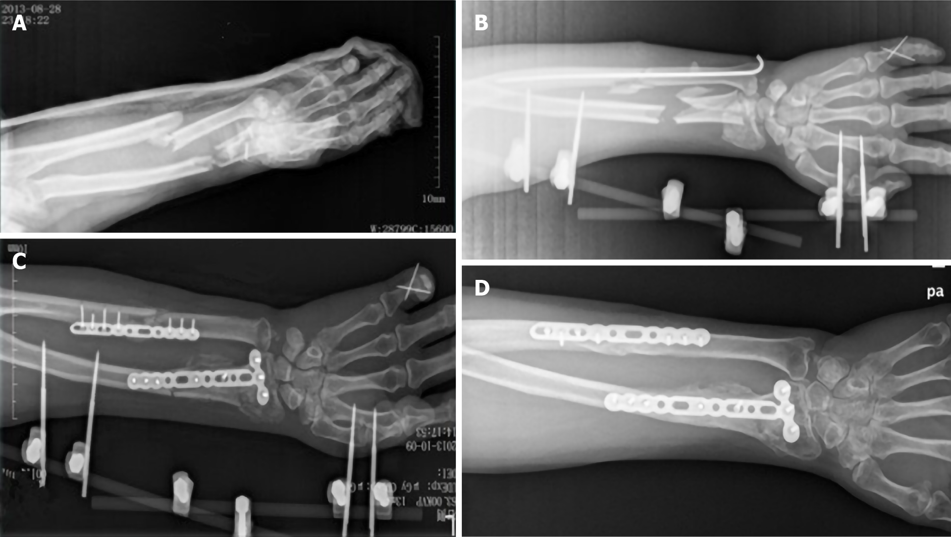
Figure 1 X-ray radiography.
A: The same day after a motorcycle accident; B: One day after the first operation; C: Twenty-eight days after the second operation; D: One year after the operation.
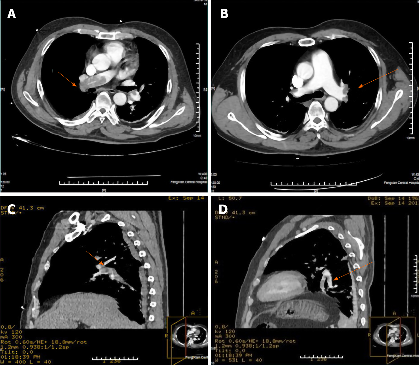
Figure 2 Computed tomographic pulmonary angiography at the onset of pulmonary embolism.
A: Horizontal plane showed filling defect in the right pulmonary artery; B: Horizontal plane showed filling defect in the left and right pulmonary arteries; C: Sagittal plane showed filling defect in the right pulmonary artery and its branches; D: Sagittal plane showed filling defect in the left pulmonary artery and its branches.
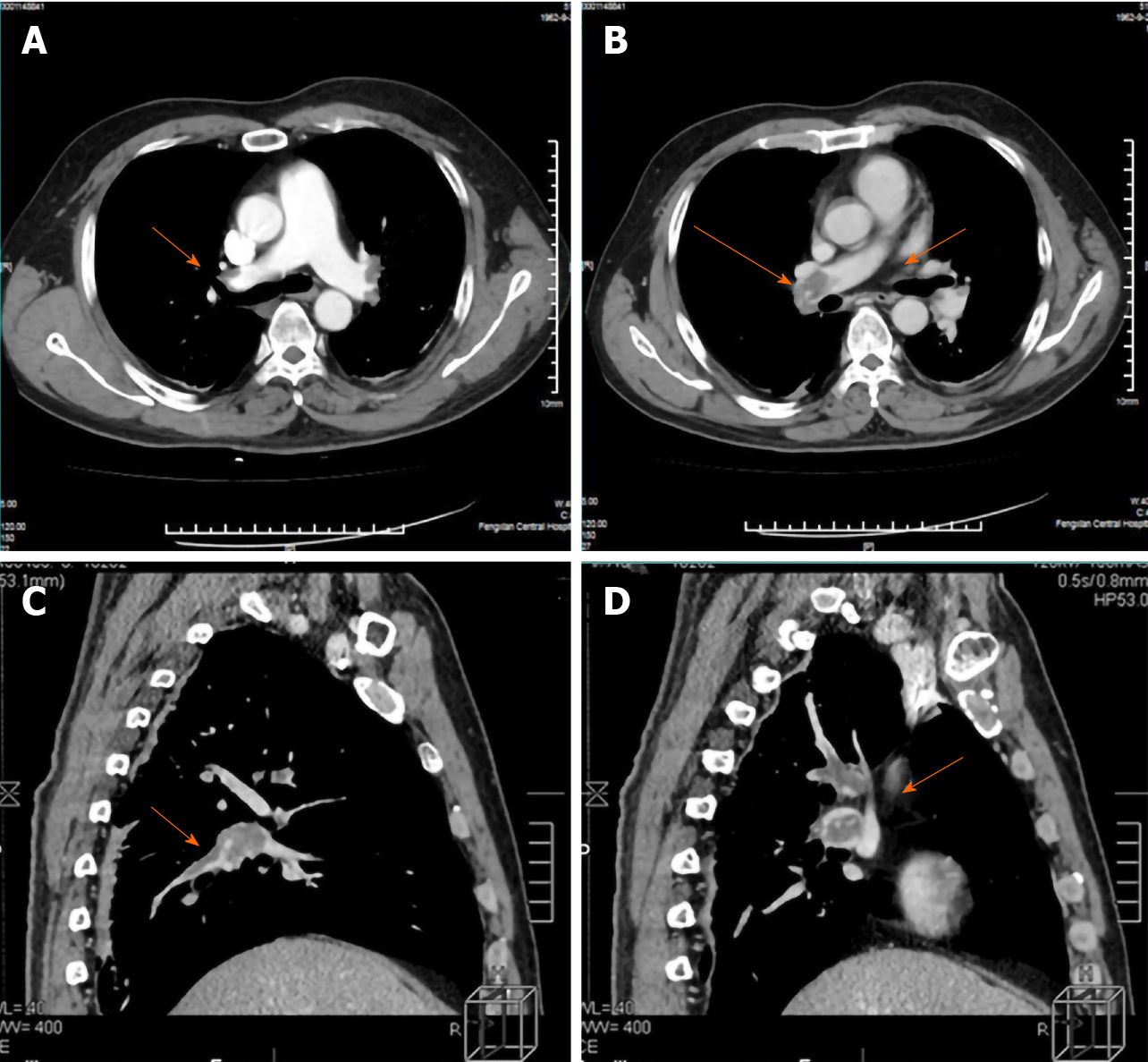
Figure 3 The ninth day after pulmonary thromboembolism, computed tomographic pulmonary angiography showed that the thrombus in both sides of the pulmonary artery did not significantly reduce.
A: Horizontal plane showed filling defect in the left and right pulmonary arteries; B: Horizontal plane showed filling defect in the right pulmonary artery; C: Sagittal plane showed filling defect in the right pulmonary artery and its branches; D: Sagittal plane showed filling defect in the left pulmonary artery and its branches.
- Citation: Lv B, Xue F, Shen YC, Hu FB, Pan MM. Pulmonary thromboembolism after distal ulna and radius fractures surgery: A case report and a literature review. World J Clin Cases 2021; 9(1): 197-203
- URL: https://www.wjgnet.com/2307-8960/full/v9/i1/197.htm
- DOI: https://dx.doi.org/10.12998/wjcc.v9.i1.197


