©The Author(s) 2020.
World J Clin Cases. Dec 26, 2020; 8(24): 6444-6449
Published online Dec 26, 2020. doi: 10.12998/wjcc.v8.i24.6444
Published online Dec 26, 2020. doi: 10.12998/wjcc.v8.i24.6444
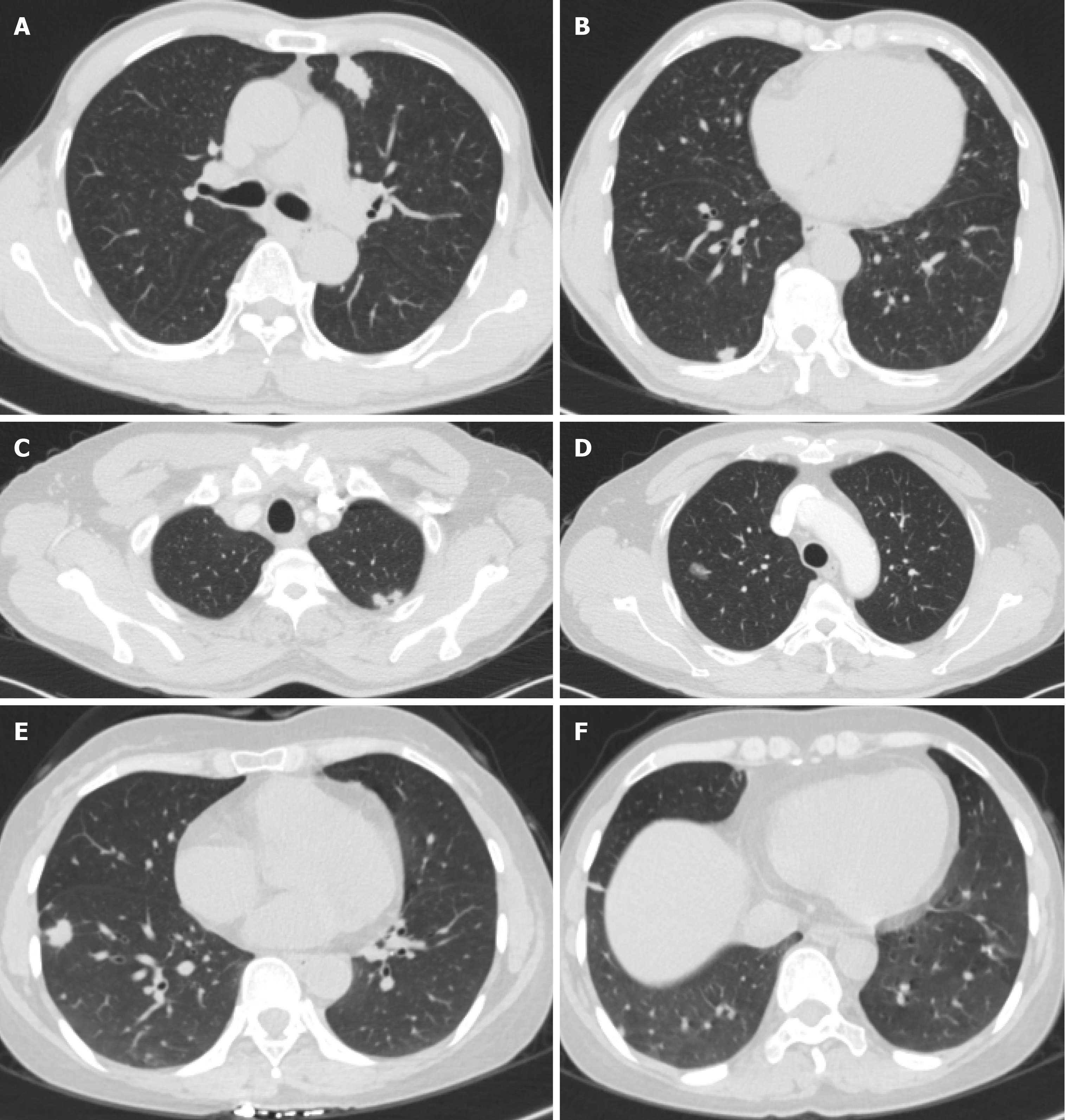
Figure 1 Computed tomography images.
A and B: Case 1 with multiple nodules in both lungs. C and D: Case 2 with a ground-glass nodule in the posterior segment of the right upper lobe apex. E and F: Case 3 with two nodules in the right lower lobe, one in the dorsal segment and the other in the outer basal segment.
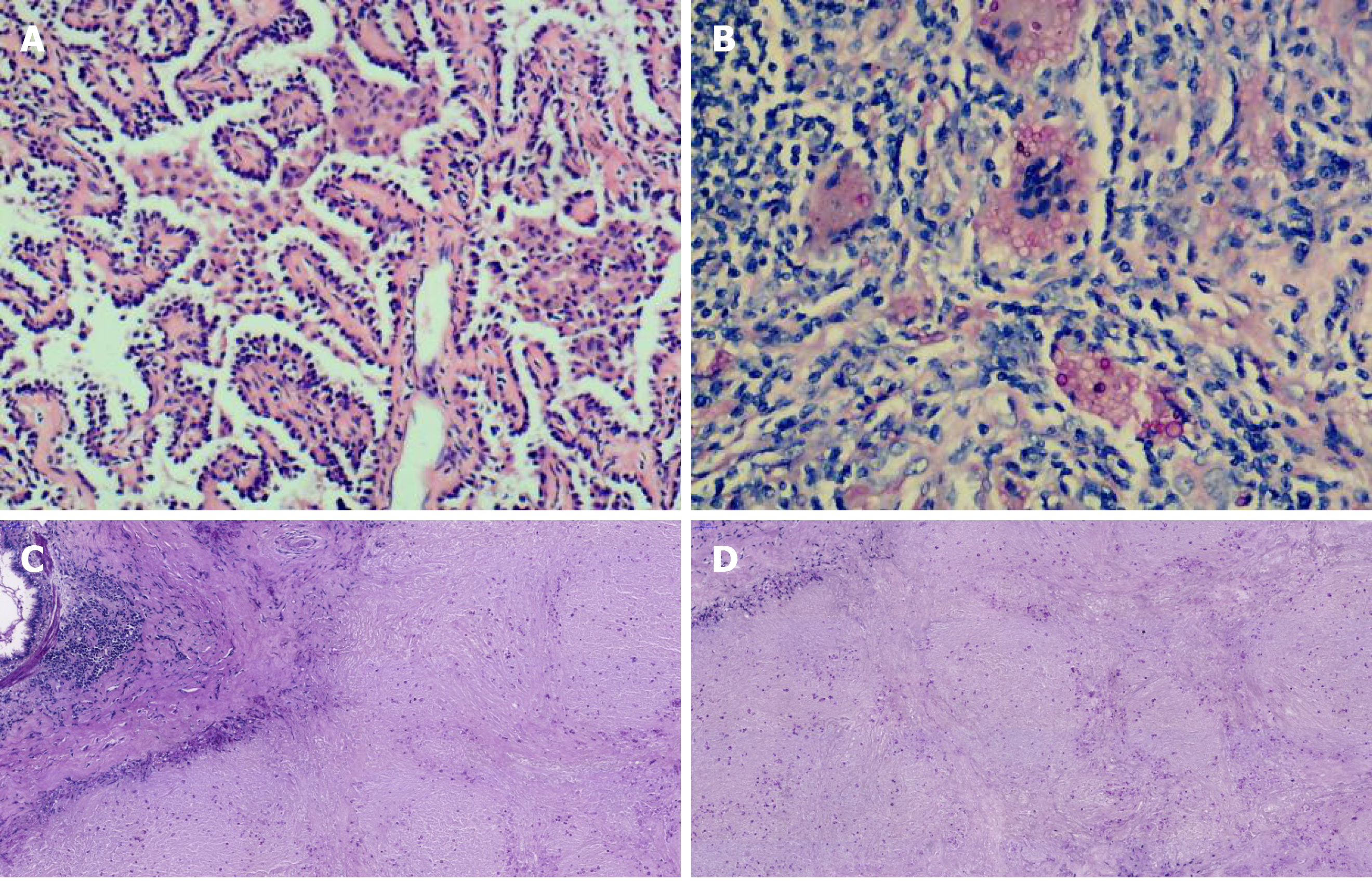
Figure 2 Histologic examination images.
A: Histopathological examination of case 1 showed that the adenocarcinoma was arranged along the alveolar septum in a papillary pattern [hematoxylin-eosin (HE) staining, original magnification × 100]; B: The yeast form of Cryptococcus neoformans was observed throughout the granulation tissue, with macrophage phagocytosis (HE staining, original magnification × 200); C: Periodic acid-Schiff (PAS) staining; D: PAS diastase staining.
- Citation: Zheng GX, Tang HJ, Huang ZP, Pan HL, Wei HY, Bai J. Clinical characteristics of pulmonary cryptococcosis coexisting with lung adenocarcinoma: Three case reports. World J Clin Cases 2020; 8(24): 6444-6449
- URL: https://www.wjgnet.com/2307-8960/full/v8/i24/6444.htm
- DOI: https://dx.doi.org/10.12998/wjcc.v8.i24.6444


