Copyright
©The Author(s) 2020.
World J Clin Cases. Nov 26, 2020; 8(22): 5821-5830
Published online Nov 26, 2020. doi: 10.12998/wjcc.v8.i22.5821
Published online Nov 26, 2020. doi: 10.12998/wjcc.v8.i22.5821
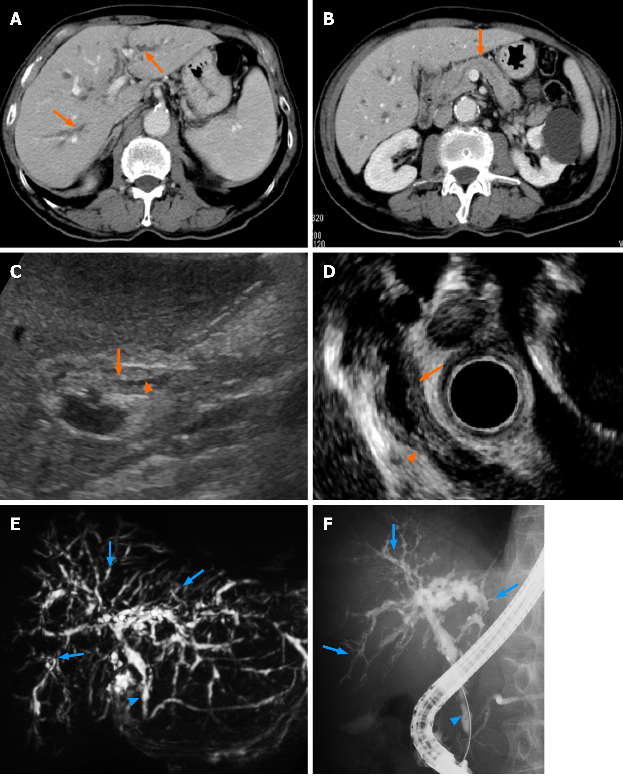
Figure 1 Contrast-enhanced computed tomography showed stricture and mild dilatation of intrahepatic bile ducts and its wall thickening with an enhance effect (A, orange arrows).
No significant swelling of the pancreas and the dilatation of main pancreatic duct were observed (B, orange arrow); C: Abdominal ultrasonography and D: Endoscopic ultrasonography showed thickening of the bile duct wall (C and D, orange arrows) and stenosis of the bile duct (C and D, orange arrowheads) in the lower bile duct. Magnetic resonance cholangiopancreatography and endoscopic retrograde cholangiopancreatography revealed stenosis of the lower bile duct (E and F, blue arrowheads) and intrahepatic bile ducts (E and F, blue arrows).
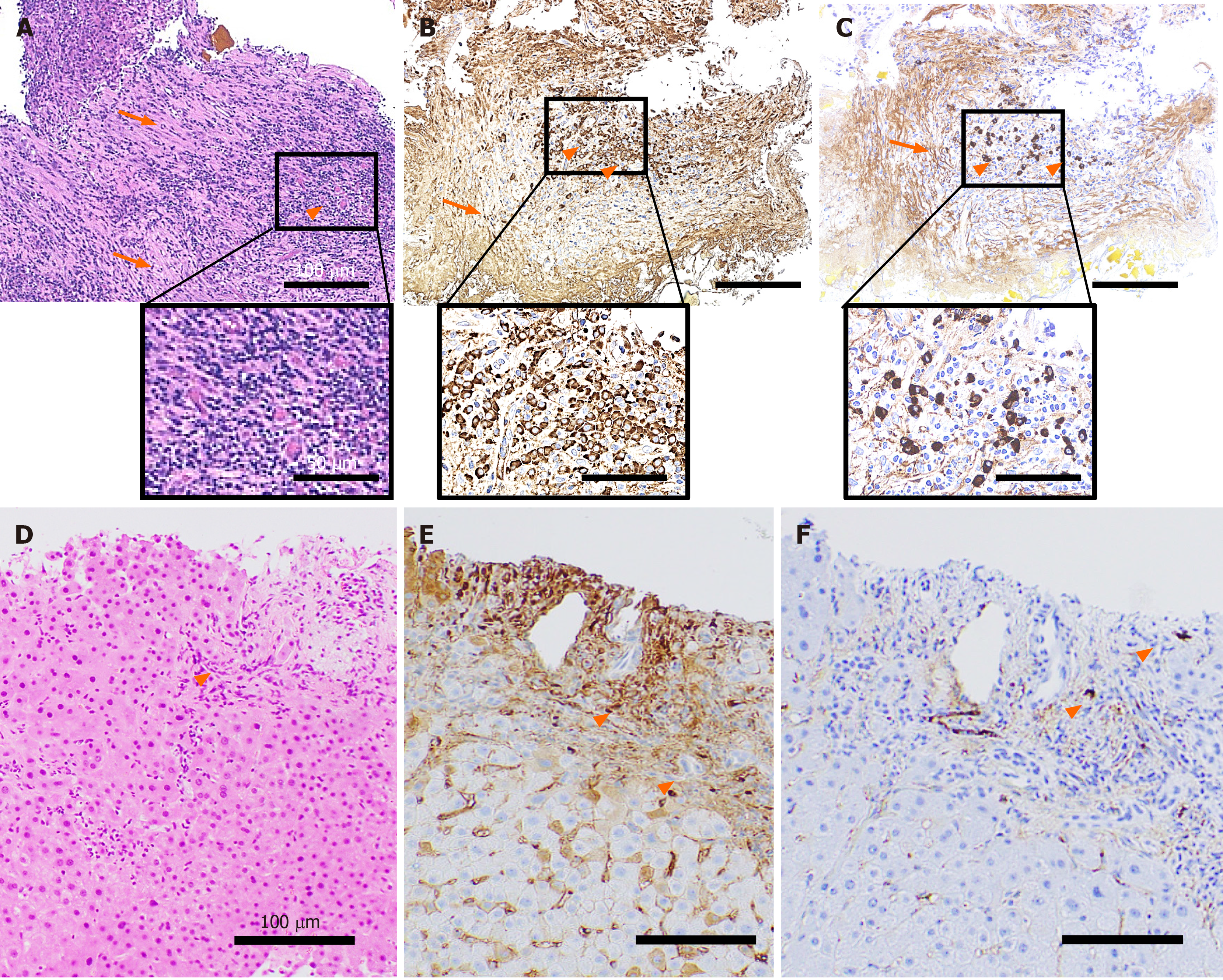
Figure 2 Histopathological findings.
A tissue sample was collected from the stenotic lower bile duct and stained with hematoxylin and eosin staining (A), IgG (B), IgG4 (C). Marked infiltration of the inflammatory cells (A-C, orange arrowheads) and storiform fibrosis (A-C, orange arrows) were observed. An increase in the number of IgG- (B) and IgG4-positive cells (C) was noted. Liver tissue showed infiltration of inflammatory cells (D: hematoxylin-eosin staining; E: IgG; F: IgG4, orange arrowheads) partly positive for IgG (E) and IgG4 (F). The scale bars represent 100 µm and 50 µm in the insets.
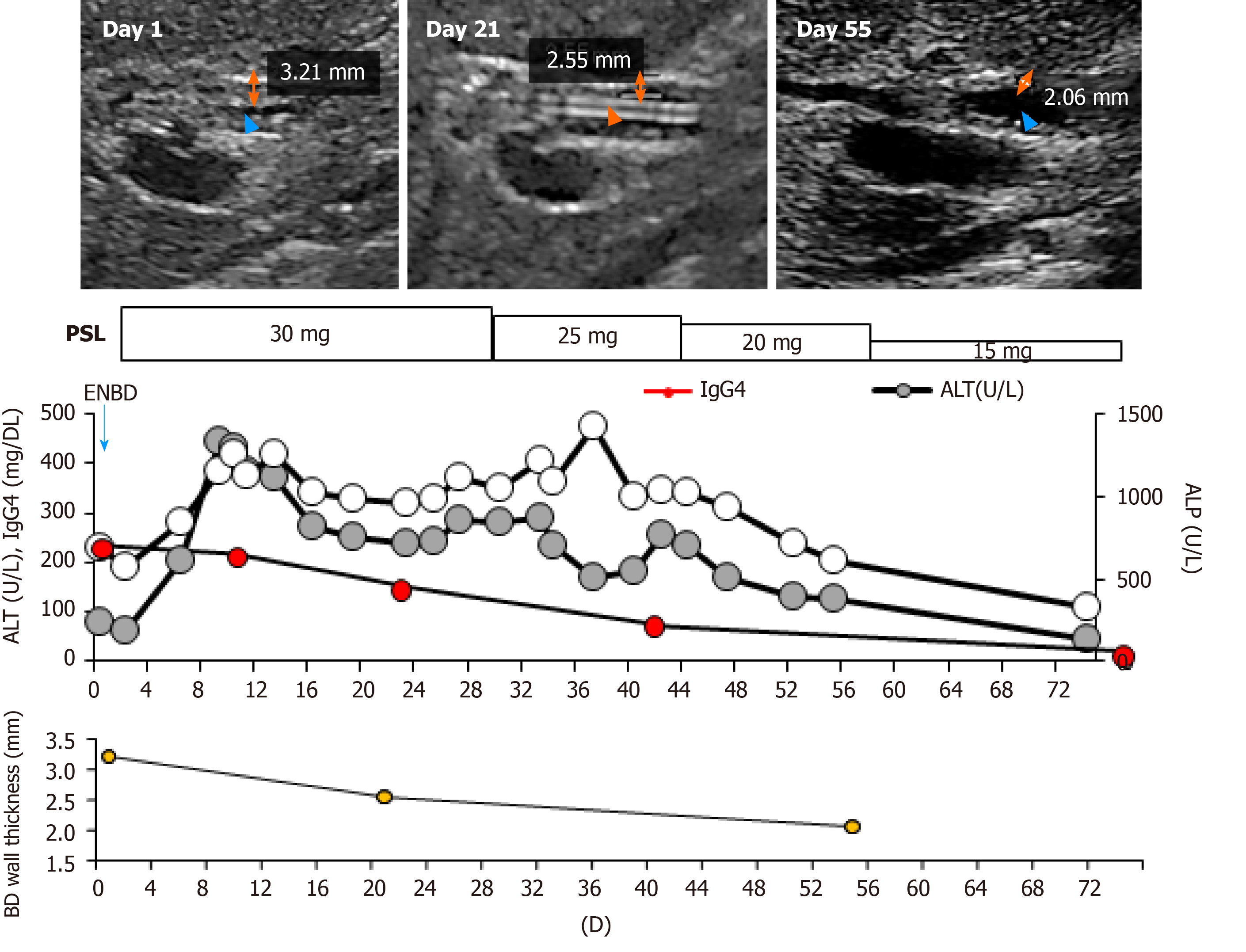
Figure 3 Clinical course.
The orange two-direction arrows indicate the wall thickness determined by abdominal ultrasonography. Orange arrowheads indicate the bile duct. The blue arrowhead indicates the endoscopic nasobiliary drainage tube. BD: Bile duct; PSL: Prednisolone; ENBD: Endoscopic nasobiliary drainage; IgG4: Immunoglobulin G4; ALT: Alanine aminotransferase; ALP: Alkaline phosphatase.
- Citation: Tanaka Y, Kamimura K, Nakamura R, Ohkoshi-Yamada M, Koseki Y, Mizusawa T, Ikarashi S, Hayashi K, Sato H, Sakamaki A, Yokoyama J, Terai S. Usefulness of ultrasonography to assess the response to steroidal therapy for the rare case of type 2b immunoglobulin G4-related sclerosing cholangitis without pancreatitis: A case report. World J Clin Cases 2020; 8(22): 5821-5830
- URL: https://www.wjgnet.com/2307-8960/full/v8/i22/5821.htm
- DOI: https://dx.doi.org/10.12998/wjcc.v8.i22.5821


