Copyright
©The Author(s) 2020.
World J Clin Cases. Nov 26, 2020; 8(22): 5795-5801
Published online Nov 26, 2020. doi: 10.12998/wjcc.v8.i22.5795
Published online Nov 26, 2020. doi: 10.12998/wjcc.v8.i22.5795
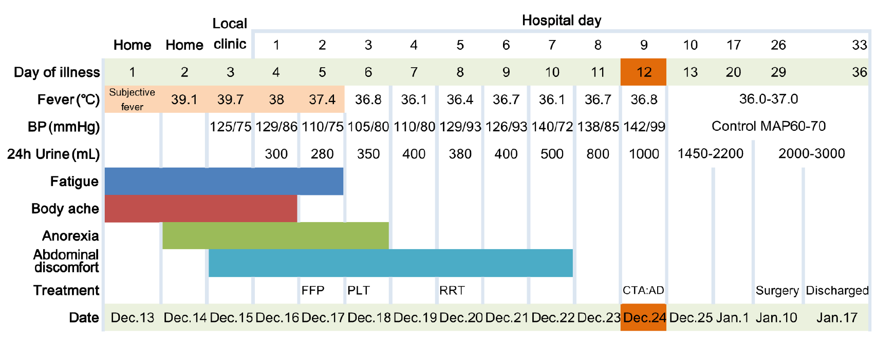
Figure 1 The patient’s manifestation and main treatment according to day of illness and hospitalization, December 13, 2018 to January 17, 2019.
AD: Aortic dissection; BP: Blood pressure; CTA: Computed tomography angiography; FFP: Fresh frozen plasma; MAP: Mean atrial blood pressure; PLT: Platelets; RRT: Renal replacement therapy.
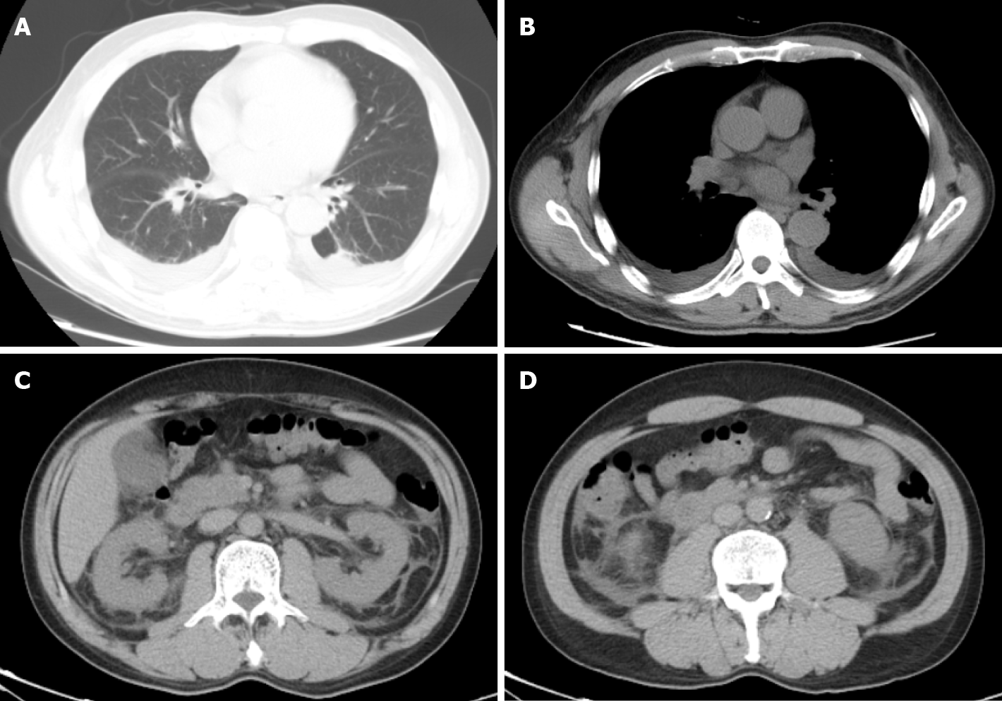
Figure 2 The computed tomography scan of chest and abdomen showed pleural effusion, perinephric effusion extended to paracolic sulcus, and slight peritoneal and pelvic effusion.
A and B: Computed tomography of the thorax and abdomen on hospital day 1 showing pleural effusion; C and D: Perinephric effusion extended to paracolic sulcus and slight peritoneal and pelvic effusion.
Figure 3 Computed tomography angiography.
A: Computed tomography angiography of the aorta on hospital day 9 showed an aortic dissection involving the left subclavian artery; B and C: Descending aorta; D: Extending to iliac artery; E: Aortic angiography during thoracic endovascular aortic repair surgery showing false lumen and true lumen (orange arrow); F: Stent graft was implanted into the vascular (orange arrow). FL: False lumen; TL: True lumen.
- Citation: Qiu FQ, Li CC, Zhou JY. Hemorrhagic fever with renal syndrome complicated with aortic dissection: A case report. World J Clin Cases 2020; 8(22): 5795-5801
- URL: https://www.wjgnet.com/2307-8960/full/v8/i22/5795.htm
- DOI: https://dx.doi.org/10.12998/wjcc.v8.i22.5795


