Copyright
©The Author(s) 2020.
World J Clin Cases. Nov 26, 2020; 8(22): 5645-5656
Published online Nov 26, 2020. doi: 10.12998/wjcc.v8.i22.5645
Published online Nov 26, 2020. doi: 10.12998/wjcc.v8.i22.5645
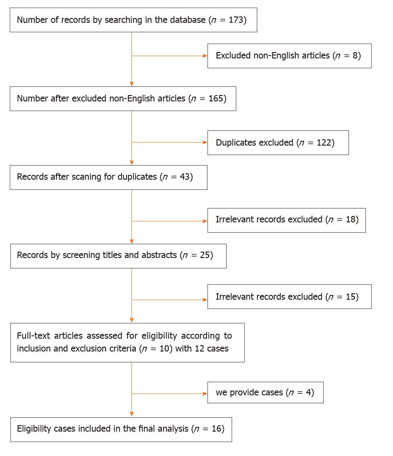
Figure 1 Flow chart of screening process.
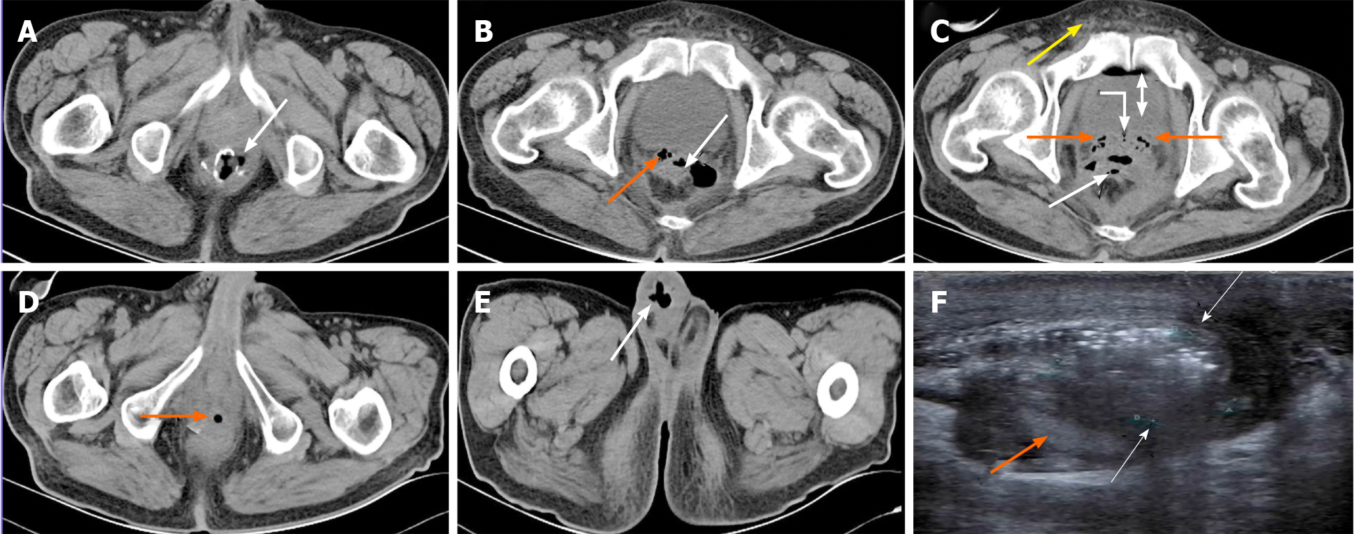
Figure 2 Imaging findings of case 1.
A-C: X-ray computed tomography (CT) demonstrated edematous rectal wall and double seminal vesicles (SV), incomplete anastomosis with an obvious rectal anastomotic leakage (AL) (white arrows). A large pelvic cavity around the AL formed a sinus from the rectum to SV, and air bubbles entered bilateral SVs (orange arrows), and ampulla of deferent duct (curved arrow). Some bubbles present in the bladder (double arrow)and a few in the right deferent duct within the spermatic cord (yellow arrow); D: CT displayed air bubbles (orange arrow) located within ejaculatory duct opening of the urethral prostate; E: Bubbles (white arrow) from the right sperm duct retrogradely entered the right edematous and infected scrotum; F: Urinary system color Doppler ultrasound showed a right epididymis tail 2.2 cm x 1.8 cm x 1.5 cm enclosed abscess and another small abscess in the right edematous scrotum with a blurred boundary, a number of strong gas echoes and peripheral rich blood (white arrows) around the testis and epididymis (orange arrow).
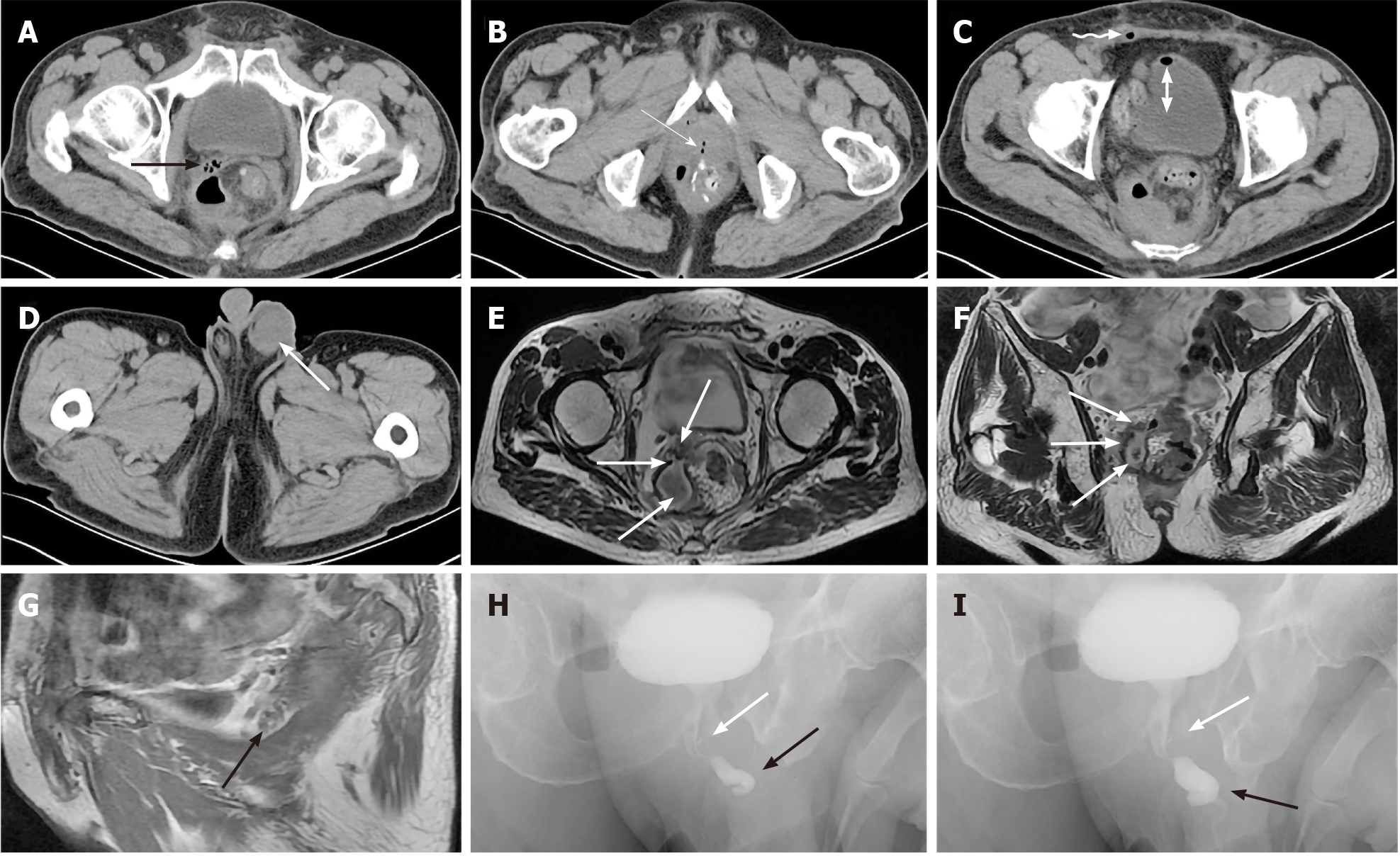
Figure 3 Imaging findings of case 2.
A-C: Computed tomography demonstrates an encapsulated cavity filled with air bubbles, pus and effusion with an air-water level connecting with the right seminal vesicles (SV) (black arrow) and ejaculatory duct opening of the urethral prostate caruncle (white arrow) after anastomotic leakage (AL). Some bubbles in the right deferent duct within the spermatic cord (curved arrow) and some in the bladder (double arrow); D: Left edematous and infective scrotum (white arrow); E, F: Cross-section and coronal plane magnetic resonance imaging (MRI) displayed a sinus between encapsulated pus cavity and right edematous SV (white arrows) secondary to AL; G: Sagittal plane MRI demonstrated some bubbles in the right edematous SV (black arrow) between bladder and rectum; H, I: Urethral retrograde radiography showed that the bladder was well filled, the anterior urethra (black arrows) was normal, the posterior urethra was slightly narrow, and the ejaculatory duct was filled with a small amount of contrast agent (white arrows).
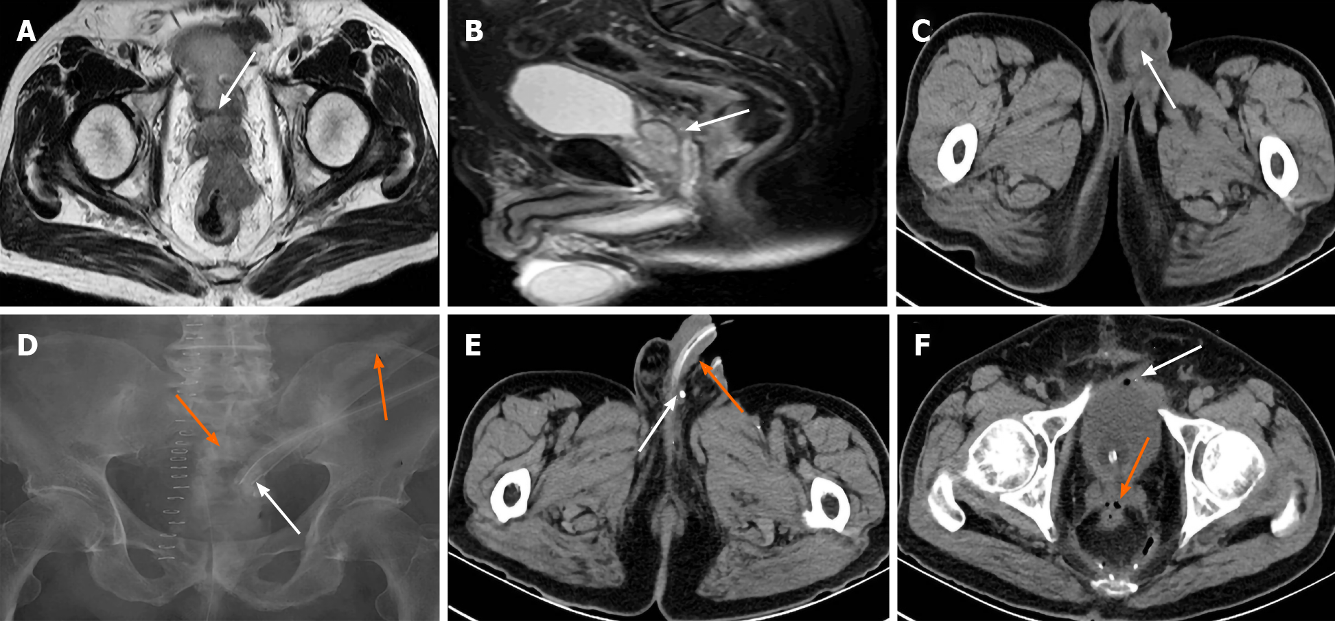
Figure 4 Imaging findings of case 3.
A, B: Cross-section and sagittal plane magnetic resonance imaging (MRI) showed that the bladder was filled well, coating of the seminal vesicles (SV) were complete, the huge rectal tumor invaded Denonvilliers’ fascia (white arrows) at the level of the SVs; C: The left swollen and infective scrotum (white arrow) by computed tomography (CT); D: Transabdominal sinus radiography showed contrast agent entering the rectum and proximal colon (orange arrows) through intrapelvic drainage tube (white arrow); E: Contrast agent residue (white arrow) in the ductus deferens beside the entrance to the epididymis but not urethra (orange arrow) by CT; F: A few effusion and bubbles in the pelvic cavity, some bubbles entering the SV (orange arrow) and bladder (white arrow) via the sinus from rectal anastomotic leakage.
Figure 5 Imaging findings of case 4.
A, B: A sinus from the rectal anastomotic site to the right seminal vesicles (SV) secondary to anastomotic leakage and air bubbles squeezed in the right SV (orange arrows) by enhanced computed tomography.
- Citation: Xia ZX, Cong JC, Zhang H. Rectoseminal vesicle fistula after radical surgery for rectal cancer: Four case reports and a literature review. World J Clin Cases 2020; 8(22): 5645-5656
- URL: https://www.wjgnet.com/2307-8960/full/v8/i22/5645.htm
- DOI: https://dx.doi.org/10.12998/wjcc.v8.i22.5645


