©The Author(s) 2020.
World J Clin Cases. Nov 6, 2020; 8(21): 5474-5479
Published online Nov 6, 2020. doi: 10.12998/wjcc.v8.i21.5474
Published online Nov 6, 2020. doi: 10.12998/wjcc.v8.i21.5474
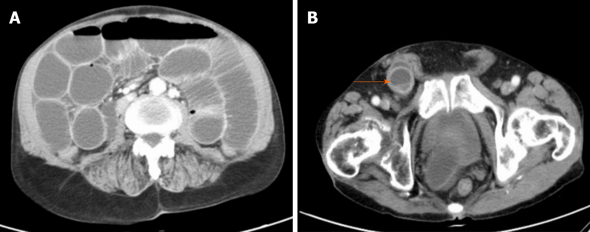
Figure 1 Computerized tomography images of the patient’s abdomen.
A: Intestinal obstruction was observed; B: A mass was observed in the right inguinal region (arrow).
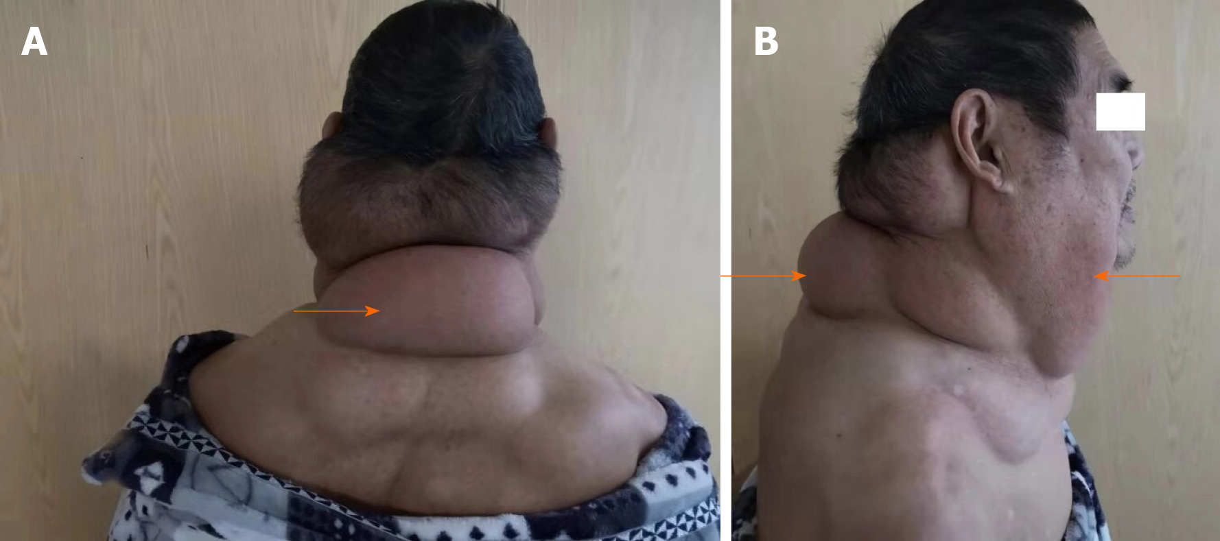
Figure 2 Photos of the patient showing a full-neck enlargement gradually developed from an egg-sized neck mass occurring 15 years ago.
A: Posterior view; B: Side view. Arrows show the neck enlargement.
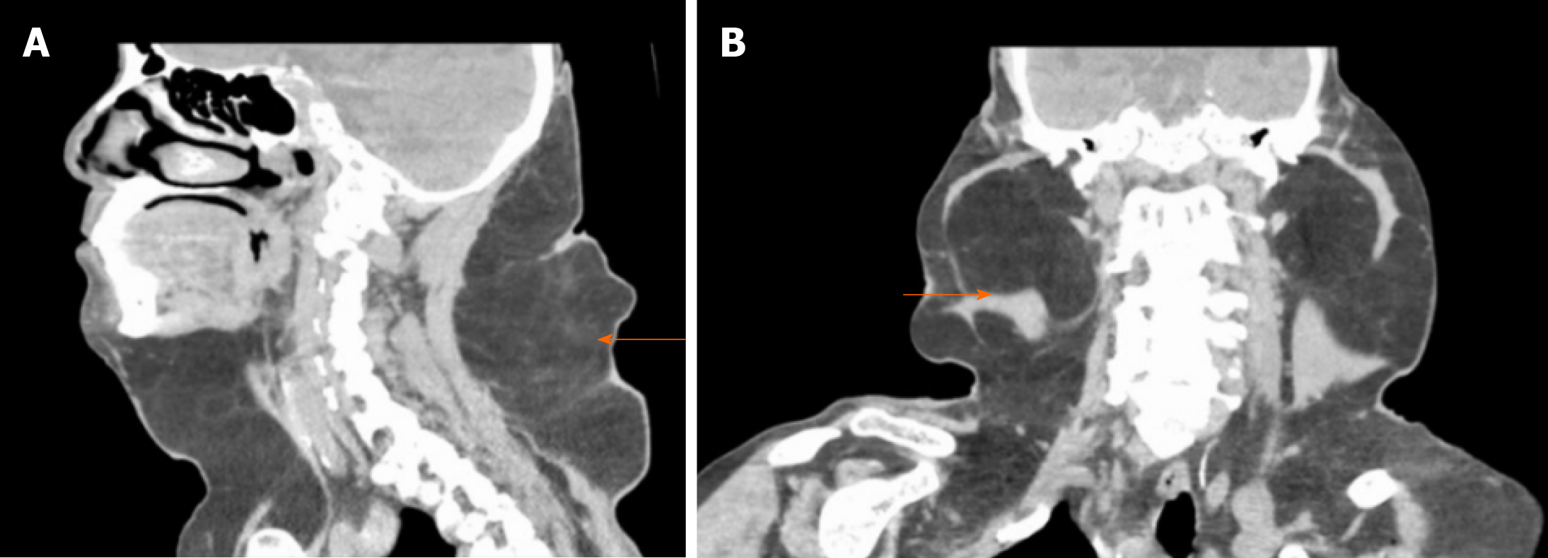
Figure 3 Computerized tomography images showing symmetrical fat accumulation in the neck and shoulder on both sides of the body.
A: An image showing fat accumulation on the front and posterior sides of the patient’s neck; B: An image showing fat accumulation on the left and right sides of the patient’s neck. Arrows show fat accumulation.
Figure 4 Computerized tomography images confirmed no recurrence of the right inguinal femoral hernia after a 1-year follow-up.
A: No intestinal protrusion in the right inguinal area; B: No obvious gas accumulation in the intestinal canal.
- Citation: Li B, Rang ZX, Weng JC, Xiong GZ, Dai XP. Benign symmetric lipomatosis (Madelung’s disease) with concomitant incarcerated femoral hernia: A case report. World J Clin Cases 2020; 8(21): 5474-5479
- URL: https://www.wjgnet.com/2307-8960/full/v8/i21/5474.htm
- DOI: https://dx.doi.org/10.12998/wjcc.v8.i21.5474


