©The Author(s) 2020.
World J Clin Cases. Nov 6, 2020; 8(21): 5457-5466
Published online Nov 6, 2020. doi: 10.12998/wjcc.v8.i21.5457
Published online Nov 6, 2020. doi: 10.12998/wjcc.v8.i21.5457
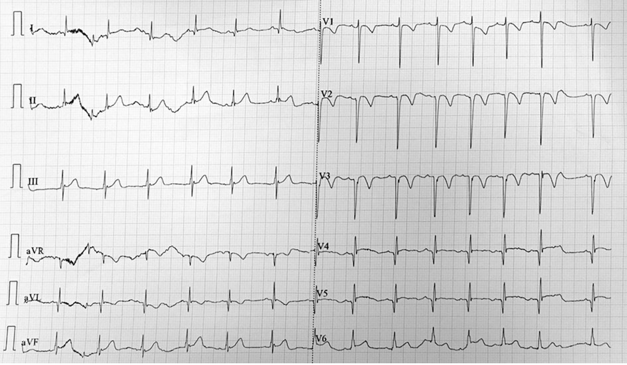
Figure 1 Electrocardiogram after cardiopulmonary resuscitation showed sinus tachycardia.
Q wave was observed in V2-4.
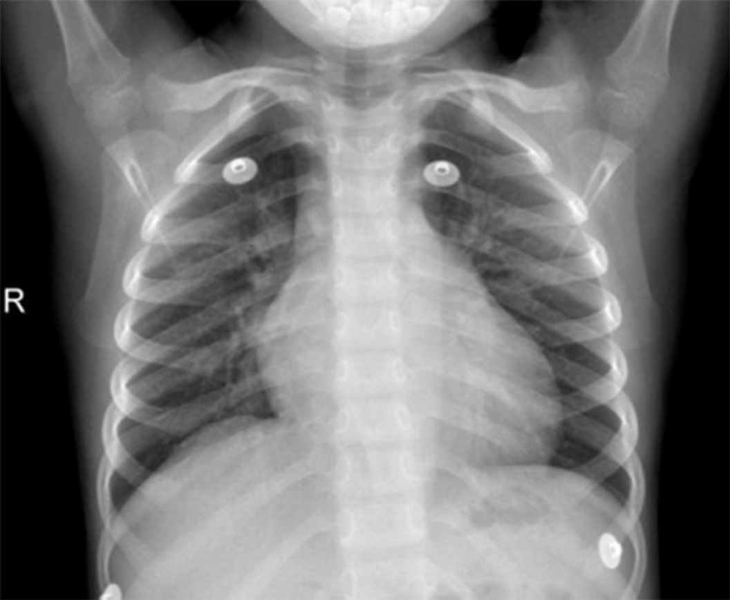
Figure 2 Chest X-ray showed enlarged heart and bulging pulmonary artery segment.
Figure 3 Transthoracic echocardiogram and 3-dimensional transthoracic echocardiogram.
Giant dilatation of the left anterior descending coronary artery (16 mm) with a massive intraluminal thrombus, dilated right coronary artery (6 mm), and enlarged left ventricle with abnormal wall motion (left ventricular ejection fraction: 39.5%) were revealed. AO: Aorta; LA: Left atrium; LAD: Left anterior descending coronary artery; LV: Left ventricle; RA: Right atrium; RCA: Right coronary artery; RV: Right ventricle.
Figure 4 Coronary computed tomography angiography 6 mo after diagnosis.
Figure 5 Cardiac magnetic resonance imaging.
Figure 6 Positron emission tomography.
A large myocardial perfusion defect in left the ventricular apical segment and survival of some mid-anterior myocardial cells were revealed.
Figure 7 Coronary angiography.
A large coronary aneurysm with a diameter of 21.5 mm × 19.7 mm at the end of the left main trunk was revealed. The filling defects in the aneurysm were considered to be thromboses. The left anterior descending branch at the distal end of the tumor was not visualized and considered to be chronic occlusion and left circumflex artery representing the tumor, blood flow, diameter, and branch without stenosis or expansion. The opening of the right coronary artery was normal. The formation of coronary aneurysms was at the beginning of the opening (12.3 mm × 6 mm) at the distal end of the tumor. Diameter and flow were normal.
- Citation: Wang T, Wang C, Zhou KY, Wang XQ, Hu N, Hua YM. Incomplete Kawasaki disease complicated with acute abdomen: A case report. World J Clin Cases 2020; 8(21): 5457-5466
- URL: https://www.wjgnet.com/2307-8960/full/v8/i21/5457.htm
- DOI: https://dx.doi.org/10.12998/wjcc.v8.i21.5457


